

Full Citation: Fluorescence Analysis of Thermoresponsive Polymers. A.G. Ryder and C. Morris, Reviews in Fluorescence 2015 ,
Annual Volumes, Vol. 8, pp. 97-126, (2015). ISBN: 978-3-319-24607-9 (Hardcover), 978-3-319-24609-3 (ebook) Springer.
Page 17 of 31
that the coupling of interfacial interaction and polymer chain dynamics causes a greater slowing of
probe diffusion. The sensitivity of the FCS methodology is very beneficial here because it allows the
differentiation of many different effects.
Diffusion of large particles formed from thermoresponsive polymers has also been studied using
more conventional fluorescence microscopy techniques like Fluorescence Recovery after
Photobleaching (FRAP). In the FRAP technique the sample is observed under a microscope (usually
a confocal laser scanning microscope), a region of interest (RoI) is rapidly bleached using relatively
high power excitation, then the region is imaged over time to determine how long it takes for new
fluorescent molecules/particles to diffuse back into the RoI. Relatively simple image analysis can be
used then to recover useful information such as diffusion rates, which in turn can be used to ascertain
changes in particle size. For thermoresponsive polymers one can use the FRAP method to observe
and measure physical effects on the micro to macro size scale. One example used FRAP to quantify
the variation in permeability of the walls of thermoresponsive hollow capsules with temperature [116].
The two di-block copolymers of PNIPAm studied were prepared as hollow capsules, and using
standard confocal laser scanning microscopy one could monitor temperature induced changes in
particle size. The permeability of the thermoresponsive shells could be assessed by comparing the
degree of infiltration of two differently sized fluorophores, 6-carboxyfluorescein and fluoresceinlabeled dextran.
ANS (1-anilino-8-naphthalene sulfonate) is one of the most widely used polarity probes because
it is highly fluorescent in low polarity solvents but is weakly fluorescent in aqueous solution [101,117].
This important feature enables one to visualize the hydrophobic regions of a given system with
minimal influence from ANS molecules remaining in the aqueous environment, and thus this
fluorophore has found widespread application in biological and material sciences [118-123]. Kujawa
et al. utilized ANS to study the concentration (0.02 to 10 g/L range) and temperature dependent
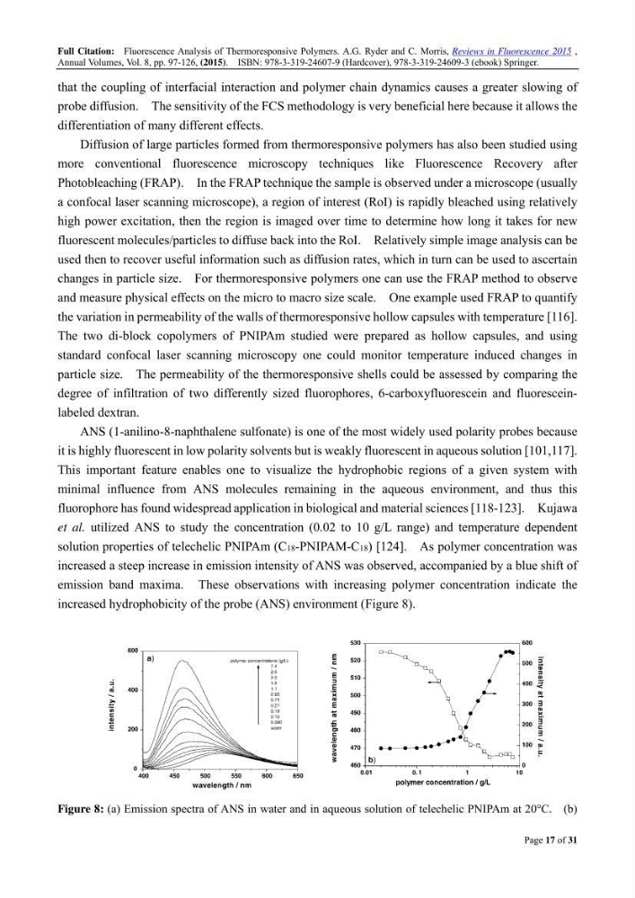
solution properties of telechelic PNIPAm (C18-PNIPAM-C18) [124]. As polymer concentration was
increased a steep increase in emission intensity of ANS was observed, accompanied by a blue shift of
emission band maxima. These observations with increasing polymer concentration indicate the
increased hydrophobicity of the probe (ANS) environment (Figure 8).
Figure 8: (a) Emission spectra of ANS in water and in aqueous solution of telechelic PNIPAm at 20C. (b)
Annual Volumes, Vol. 8, pp. 97-126, (2015). ISBN: 978-3-319-24607-9 (Hardcover), 978-3-319-24609-3 (ebook) Springer.
Page 17 of 31
that the coupling of interfacial interaction and polymer chain dynamics causes a greater slowing of
probe diffusion. The sensitivity of the FCS methodology is very beneficial here because it allows the
differentiation of many different effects.
Diffusion of large particles formed from thermoresponsive polymers has also been studied using
more conventional fluorescence microscopy techniques like Fluorescence Recovery after
Photobleaching (FRAP). In the FRAP technique the sample is observed under a microscope (usually
a confocal laser scanning microscope), a region of interest (RoI) is rapidly bleached using relatively
high power excitation, then the region is imaged over time to determine how long it takes for new
fluorescent molecules/particles to diffuse back into the RoI. Relatively simple image analysis can be
used then to recover useful information such as diffusion rates, which in turn can be used to ascertain
changes in particle size. For thermoresponsive polymers one can use the FRAP method to observe
and measure physical effects on the micro to macro size scale. One example used FRAP to quantify
the variation in permeability of the walls of thermoresponsive hollow capsules with temperature [116].
The two di-block copolymers of PNIPAm studied were prepared as hollow capsules, and using
standard confocal laser scanning microscopy one could monitor temperature induced changes in
particle size. The permeability of the thermoresponsive shells could be assessed by comparing the
degree of infiltration of two differently sized fluorophores, 6-carboxyfluorescein and fluoresceinlabeled dextran.
ANS (1-anilino-8-naphthalene sulfonate) is one of the most widely used polarity probes because
it is highly fluorescent in low polarity solvents but is weakly fluorescent in aqueous solution [101,117].
This important feature enables one to visualize the hydrophobic regions of a given system with
minimal influence from ANS molecules remaining in the aqueous environment, and thus this
fluorophore has found widespread application in biological and material sciences [118-123]. Kujawa
et al. utilized ANS to study the concentration (0.02 to 10 g/L range) and temperature dependent
solution properties of telechelic PNIPAm (C18-PNIPAM-C18) [124]. As polymer concentration was
increased a steep increase in emission intensity of ANS was observed, accompanied by a blue shift of
emission band maxima. These observations with increasing polymer concentration indicate the
increased hydrophobicity of the probe (ANS) environment (Figure 8).
Figure 8: (a) Emission spectra of ANS in water and in aqueous solution of telechelic PNIPAm at 20C. (b)