

seven were positive for V. cholerae 01 by conventional culture methods. Over 3,000 colonies were picked for slide agglutination for this determination. Using fluorescent antibody techniques, however, 51 out of the 52 samples were confirmed as positive for V.
cholerae. All samples positive by culture, were positive by staining. Also, recovery of V.
cholerae on media was higher at the earlier stages of enrichment incubation, and not
recoverable at later periods. Fluorescent antibody results remained positive throughout.
Date
Source (1)
Culture
y ibrio cholerae MPN (2)
Epifluorescent
Total (3)
01
01
6/15/81
W
24
<3.0
460
6/15/81
SD
1.1
<0.3
>110
6/23/81
Sw
11,000
16
>1100
9/14/81
W
29/4,600
<3.0
150
9/14/81
Sd
1.2/21
<0.3
4.2
9/15/81
Sw
11,000
<3.0
20
12/7/81
W
<3.0
<3.0
<3.0
12/7/81
Sd
<0.3
<0.3
<0.3
12/15/81
Sw
<3.0
<3.0
<3.0
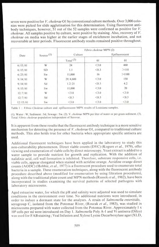
Table I : Vibrio Cholerae culture and epifluorescent MPN results of Louisiana samples.
(1). Water : W, Sediment : Sd, Sewage : Sw. (2). V. cholerae MPN per liter of water or per gram sediment. (3).
Total Vibrio cholerae population independent of Serovar.
It is apparent from these results that the fluorescent antibody technique is a more sensitive
mechanism for detecting the presence of V. cholerae 01, compared to traditional culture
methods. This also holds true for other bacteria when appropriate specific antisera are
used.
Additional fluorescent techniques have been applied in the laboratory to study this
non-culturability phenomenon. Direct viable counts (DVC) (Kogure et al., 1979), offer
viewing and enumeration of viable cells by direct microscopy. Yeast extract is added to a
water sample to provide nutrient for growth and replication. With the addition of
nalidixic acid, cell wall formation is inhibited. Therefore, substrate responsive cells, i.e.
viable cells, appear elongated when stained with acridine orange. Acridine orange direct
counts (AODC) (Hobbie, et al., 1977) is a fluorescent procedure used to enumerate total
bacteria in a sample. These enumeration techniques, along with the fluorescent antibody
procedure described above (modified for enumeration by using filtration procedures),
along with the traditional plate count and M PN methods (Russek et ai, 1982), have been
incorporated in studies examining the survival patterns of selected pathogens with
laboratory microcosms.
Aged estuarine water, for which the pH and salinity were adjusted was used to simulate
conditions in the environment over time. No additional nutrients were introduced, in
order to induce a dormant state for the analyses. A strain of Salmonella enteritidis,
serogroup C, isolated from the Potomac River, (Roszak et al., 1983), was studied in
microcosms prepared with water collected from the Potomac River. Approximately 1 x
10
5 cells per ml were introduced on Day 1. Salmonella Poly A-l and Vi antisera (Difco)
was used for FAB staining ; Veal Infusion and Xylose Lysine Decarboxylase agar (XLD,
509
cholerae. All samples positive by culture, were positive by staining. Also, recovery of V.
cholerae on media was higher at the earlier stages of enrichment incubation, and not
recoverable at later periods. Fluorescent antibody results remained positive throughout.
Date
Source (1)
Culture
y ibrio cholerae MPN (2)
Epifluorescent
Total (3)
01
01
6/15/81
W
24
<3.0
460
6/15/81
SD
1.1
<0.3
>110
6/23/81
Sw
11,000
16
>1100
9/14/81
W
29/4,600
<3.0
150
9/14/81
Sd
1.2/21
<0.3
4.2
9/15/81
Sw
11,000
<3.0
20
12/7/81
W
<3.0
<3.0
<3.0
12/7/81
Sd
<0.3
<0.3
<0.3
12/15/81
Sw
<3.0
<3.0
<3.0
Table I : Vibrio Cholerae culture and epifluorescent MPN results of Louisiana samples.
(1). Water : W, Sediment : Sd, Sewage : Sw. (2). V. cholerae MPN per liter of water or per gram sediment. (3).
Total Vibrio cholerae population independent of Serovar.
It is apparent from these results that the fluorescent antibody technique is a more sensitive
mechanism for detecting the presence of V. cholerae 01, compared to traditional culture
methods. This also holds true for other bacteria when appropriate specific antisera are
used.
Additional fluorescent techniques have been applied in the laboratory to study this
non-culturability phenomenon. Direct viable counts (DVC) (Kogure et al., 1979), offer
viewing and enumeration of viable cells by direct microscopy. Yeast extract is added to a
water sample to provide nutrient for growth and replication. With the addition of
nalidixic acid, cell wall formation is inhibited. Therefore, substrate responsive cells, i.e.
viable cells, appear elongated when stained with acridine orange. Acridine orange direct
counts (AODC) (Hobbie, et al., 1977) is a fluorescent procedure used to enumerate total
bacteria in a sample. These enumeration techniques, along with the fluorescent antibody
procedure described above (modified for enumeration by using filtration procedures),
along with the traditional plate count and M PN methods (Russek et ai, 1982), have been
incorporated in studies examining the survival patterns of selected pathogens with
laboratory microcosms.
Aged estuarine water, for which the pH and salinity were adjusted was used to simulate
conditions in the environment over time. No additional nutrients were introduced, in
order to induce a dormant state for the analyses. A strain of Salmonella enteritidis,
serogroup C, isolated from the Potomac River, (Roszak et al., 1983), was studied in
microcosms prepared with water collected from the Potomac River. Approximately 1 x
10
5 cells per ml were introduced on Day 1. Salmonella Poly A-l and Vi antisera (Difco)
was used for FAB staining ; Veal Infusion and Xylose Lysine Decarboxylase agar (XLD,
509