

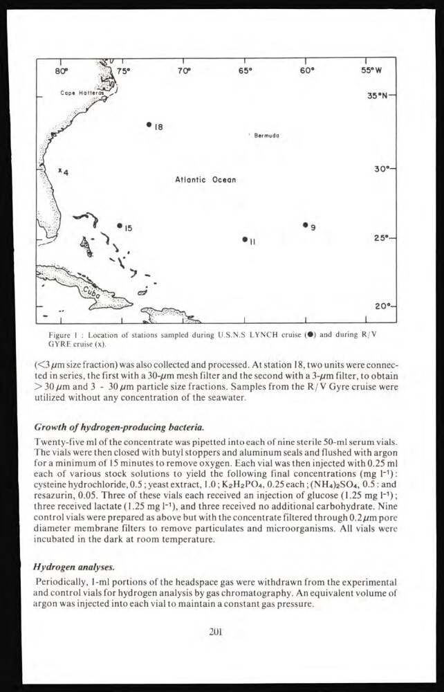
Figure I : Location of stations sampled during U.S.N.S LYNCH cruise (•) and during R/V
GYRE cruise (x).
(<3 µm size fraction) was also collected and processed. At station 18, two units were connected in series, the first with a 30-µm mesh filter and the second with a 3-µm filter, to obtain
> 30 pm and 3-30 pm particle size fractions. Samples from the R/V Gyre cruise were
utilized without any concentration of the seawater.
Growth of hydrogen-producing bacteria.
Twenty-five ml of the concentrate was pipetted into each of nine sterile 50-ml serum vials.
The vials were then closed with butyl stoppers and aluminum seals and flushed with argon
for a minimum of 15 minutes to remove oxygen. Each vial was then injected with 0.25 ml
each of various stock solutions to yield the following final concentrations (mg l -1 ):
cysteine hydrochloride, 0.5 ; yeast extract, 1.0 ; K 2 H 2 PO 4 , 0.25 each ; (NH 4 ) 2 SO 4 , 0.5 : and
resazurin, 0.05. Three of these vials each received an injection of glucose (1.25 mg l -1 );
three received lactate (1.25 mg 1 -1 ), and three received no additional carbohydrate. Nine
control vials were prepared as above but with the concentrate filtered through 0.2 µm pore
diameter membrane filters to remove particulates and microorganisms. All vials were
incubated in the dark at room temperature.
Hydrogen analyses.
Periodically, 1-ml portions of the headspace gas were withdrawn from the experimental
and control vials for hydrogen analysis by gas chromatography. An equivalent volume of
argon was injected into each vial to maintain a constant gas pressure.
201
GYRE cruise (x).
(<3 µm size fraction) was also collected and processed. At station 18, two units were connected in series, the first with a 30-µm mesh filter and the second with a 3-µm filter, to obtain
> 30 pm and 3-30 pm particle size fractions. Samples from the R/V Gyre cruise were
utilized without any concentration of the seawater.
Growth of hydrogen-producing bacteria.
Twenty-five ml of the concentrate was pipetted into each of nine sterile 50-ml serum vials.
The vials were then closed with butyl stoppers and aluminum seals and flushed with argon
for a minimum of 15 minutes to remove oxygen. Each vial was then injected with 0.25 ml
each of various stock solutions to yield the following final concentrations (mg l -1 ):
cysteine hydrochloride, 0.5 ; yeast extract, 1.0 ; K 2 H 2 PO 4 , 0.25 each ; (NH 4 ) 2 SO 4 , 0.5 : and
resazurin, 0.05. Three of these vials each received an injection of glucose (1.25 mg l -1 );
three received lactate (1.25 mg 1 -1 ), and three received no additional carbohydrate. Nine
control vials were prepared as above but with the concentrate filtered through 0.2 µm pore
diameter membrane filters to remove particulates and microorganisms. All vials were
incubated in the dark at room temperature.
Hydrogen analyses.
Periodically, 1-ml portions of the headspace gas were withdrawn from the experimental
and control vials for hydrogen analysis by gas chromatography. An equivalent volume of
argon was injected into each vial to maintain a constant gas pressure.
201