

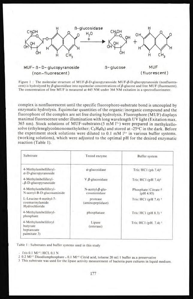
MUF- (3-D-glucopyranoside
(non-fluorescent)
(3-glucose
(fluorescent)
Figure 1 : The molecular structure of MUF-/3-D-glucopyranoside MUF-β-D-glucopyranoside (nonfluorescent) is hydrolyzed by β-glucosidase into equimolar concentrations of β-glucose and free MUF (fluorescent).
The concentration of free MUF is measured at 445 NM under 364 NM exitation in a spectrofluorimeter.
complex is nonfluorescent until the specific fluorophore-substrate bond is uncoupled by
enzymatic hydrolysis. Equimolar quantities of the organic/inorganic compound and the
fluorophore of the complex are set free during hydrolysis. Fluorophore (MUF) displays
maximal fluorescence under illumination with long wavelength UV light (Exitation max.
365 nm). Stock solutions of MUF-substrates(5 mM l
1_ ) were prepared in methylcellosolve (ethyleneglycolmonomethylether; C 3 H 8 O 2 ) and stored at -25°C in the dark. Before
the experiment stock solutions were diluted to 0.1 mM l
1- in various buffer systems,
(working solutions), which were adjusted to the optimal pH for the desired enzymatic
reaction (Table 1).
Substrate
Tested enzyme
Buffer system
4-Methylumbelliferylα-D-glucopyranoside
α-glucosidase
Tris/HCl (ph 7.4)
1
4-Methylmbelliferyl. β-D-glucopyranoside
V β-glucosidase
Tris/HCl (pH 7.4)
1
4-MethylumbelliferylN-acetyl-B-D-glucosaminide
N-acetyl-β-glucosaminidase
Phosphate/Citrate 2
(pH 4.95)
L-Leucine-4-methyl-7coumarinylamide
Hydrochloride
protease
(aminopeptidase)
Tris/HCl (pH 7.4)
1
4-Methylumbelliferylphosphate
phosphatase
Tris/HCl (pH 8.3)
1
4-Methylumbelliferyl
butyrate
heptanoate
palmitate 3)
Lipase
(esterase)
Tris/HCl (pH. 7.4)
1
Table 1 : Substrates and buffer systems used in this study
. Tris 0.1 Ml -1 /HCL 0.1 N
2 0.2 Ml -1 Disodiumphosphate + 0.1 Ml*
1 Citrid acid, toluene 20 ml/l buffer as a preservative
3 This substrate was used for the lipase activity measurement of bacteria pure cultures in liquid medium.
177
(non-fluorescent)
(3-glucose
(fluorescent)
Figure 1 : The molecular structure of MUF-/3-D-glucopyranoside MUF-β-D-glucopyranoside (nonfluorescent) is hydrolyzed by β-glucosidase into equimolar concentrations of β-glucose and free MUF (fluorescent).
The concentration of free MUF is measured at 445 NM under 364 NM exitation in a spectrofluorimeter.
complex is nonfluorescent until the specific fluorophore-substrate bond is uncoupled by
enzymatic hydrolysis. Equimolar quantities of the organic/inorganic compound and the
fluorophore of the complex are set free during hydrolysis. Fluorophore (MUF) displays
maximal fluorescence under illumination with long wavelength UV light (Exitation max.
365 nm). Stock solutions of MUF-substrates(5 mM l
1_ ) were prepared in methylcellosolve (ethyleneglycolmonomethylether; C 3 H 8 O 2 ) and stored at -25°C in the dark. Before
the experiment stock solutions were diluted to 0.1 mM l
1- in various buffer systems,
(working solutions), which were adjusted to the optimal pH for the desired enzymatic
reaction (Table 1).
Substrate
Tested enzyme
Buffer system
4-Methylumbelliferylα-D-glucopyranoside
α-glucosidase
Tris/HCl (ph 7.4)
1
4-Methylmbelliferyl. β-D-glucopyranoside
V β-glucosidase
Tris/HCl (pH 7.4)
1
4-MethylumbelliferylN-acetyl-B-D-glucosaminide
N-acetyl-β-glucosaminidase
Phosphate/Citrate 2
(pH 4.95)
L-Leucine-4-methyl-7coumarinylamide
Hydrochloride
protease
(aminopeptidase)
Tris/HCl (pH 7.4)
1
4-Methylumbelliferylphosphate
phosphatase
Tris/HCl (pH 8.3)
1
4-Methylumbelliferyl
butyrate
heptanoate
palmitate 3)
Lipase
(esterase)
Tris/HCl (pH. 7.4)
1
Table 1 : Substrates and buffer systems used in this study
. Tris 0.1 Ml -1 /HCL 0.1 N
2 0.2 Ml -1 Disodiumphosphate + 0.1 Ml*
1 Citrid acid, toluene 20 ml/l buffer as a preservative
3 This substrate was used for the lipase activity measurement of bacteria pure cultures in liquid medium.
177