

38
T. TOMITA
this is interrupted by the cornea-positive PII. The a-wave of the ERG
thus represents the early onset of PIII, while the b-wave the rise of PII.
The PI corresponds to the c-wave in the ERG, and, as earlier described,
its amplitude depends on the state of adaptation. The d-wave at the cessation of light is, according to this analysis, either a positive or negative
deflection, determined by the amplitude and time course of the off-effects
of PI1 and PI11 which are opposite in polarity. In most fishes, the PI11
predominates especially in the photopic state, resulting in a large a-wave
and a large cornea-positive d-wave.
Originally, the component analysis of the ERG was made by isolation
of one component from others using various physical and chemical agents.
In general, the PI1 is more susceptible than the PI11 to various agents.
One exception is alcohol, which results in a marked increase in the bwave and decrease in the a- and d-wave (Bernhard and Skoglund, 1941).
Granit (1947) provides evidence that the PI1 is associated with
excitatory processes in the retina and the PI11 with inhibitory. This view
is consistent with the common observation that a burst of impulse discharge in the optic nerve occurs with the b-wave, which is the rise of the
excitatory PII, and another burst with the d-wave, which is the rebound
Distance
W
in retina
2- -0 250t
Q
E
a
-250t \*/
\
- 500 1
0
\
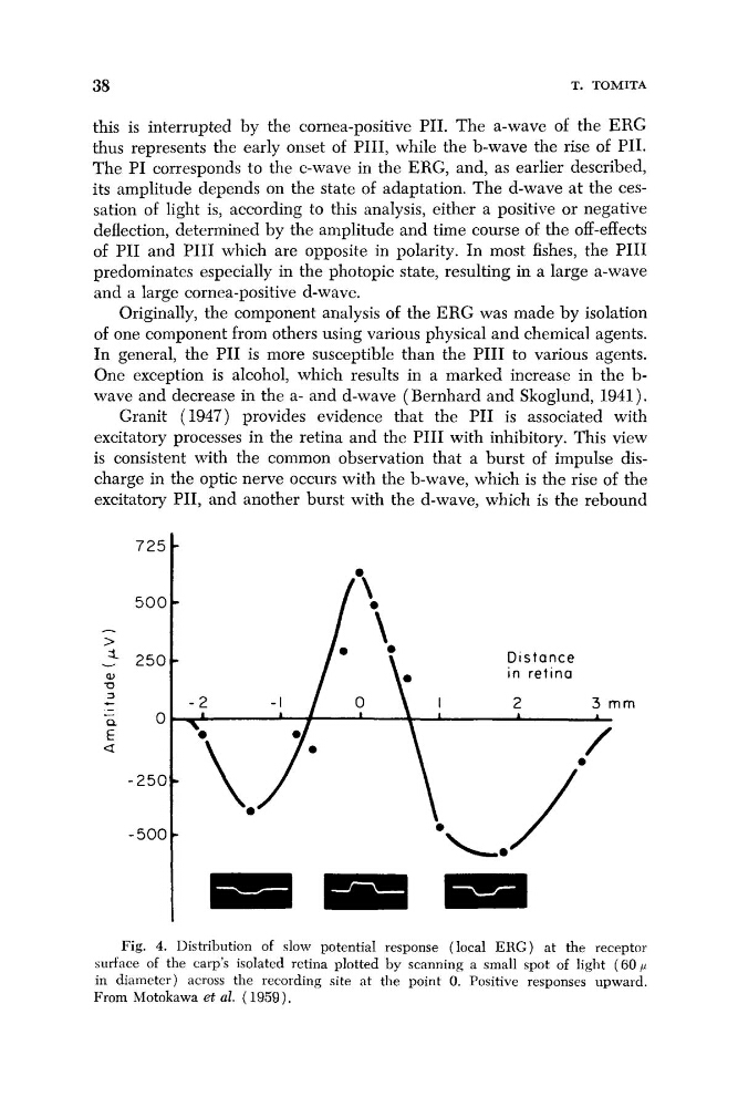
Fig. 4. Distribution of slow potential response (local ERG) at the receptor
surface of the carp’s isolated retina plotted by scanning a small spot of light (60 , U
in diameter) across the recording site at the point 0. Positive responses upward.
From Motokawa et al. ( 1959).
T. TOMITA
this is interrupted by the cornea-positive PII. The a-wave of the ERG
thus represents the early onset of PIII, while the b-wave the rise of PII.
The PI corresponds to the c-wave in the ERG, and, as earlier described,
its amplitude depends on the state of adaptation. The d-wave at the cessation of light is, according to this analysis, either a positive or negative
deflection, determined by the amplitude and time course of the off-effects
of PI1 and PI11 which are opposite in polarity. In most fishes, the PI11
predominates especially in the photopic state, resulting in a large a-wave
and a large cornea-positive d-wave.
Originally, the component analysis of the ERG was made by isolation
of one component from others using various physical and chemical agents.
In general, the PI1 is more susceptible than the PI11 to various agents.
One exception is alcohol, which results in a marked increase in the bwave and decrease in the a- and d-wave (Bernhard and Skoglund, 1941).
Granit (1947) provides evidence that the PI1 is associated with
excitatory processes in the retina and the PI11 with inhibitory. This view
is consistent with the common observation that a burst of impulse discharge in the optic nerve occurs with the b-wave, which is the rise of the
excitatory PII, and another burst with the d-wave, which is the rebound
Distance
W
in retina
2- -0 250t
Q
E
a
-250t \*/
\
- 500 1
0
\
Fig. 4. Distribution of slow potential response (local ERG) at the receptor
surface of the carp’s isolated retina plotted by scanning a small spot of light (60 , U
in diameter) across the recording site at the point 0. Positive responses upward.
From Motokawa et al. ( 1959).