

M. V. L. BENNETT
462
tween cells; a relatively more depolarized cell tends to depolarize and
excite its less depolarized neighbors and is itself made less depolarized
and inhibited by them. Thus, the neurons tend to fire synchronously. The
synapses are both excitatory and inhibitory, and it seems reasonable
to call them synchronizing synapses (Bennett, 1968b). One may ask
whether mutually excitatory, chemically transmitting synapses could also
mediate synchronization, As will be seen below synchronization in most
of the electric organ systems is so precise that the negligible delay of
electrically mediated transmission is required; the delay associated with
chemically mediated transmission would be too great.
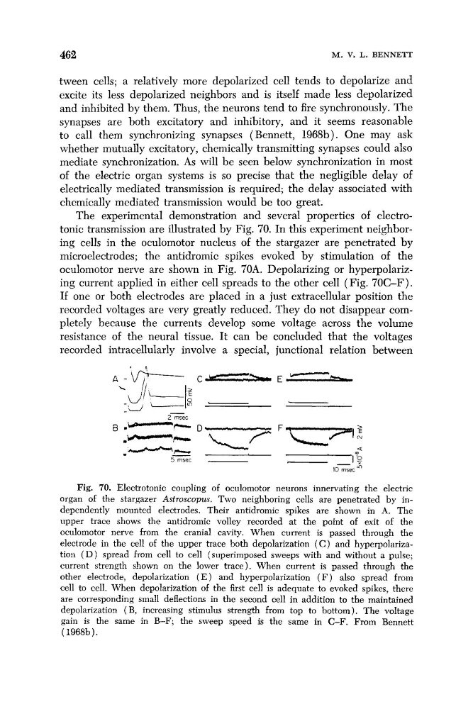
The experimental demonstration and several properties of electrotonic transmission are illustrated by Fig. 70. In this experiment neighboring cells in the oculomotor nucleus of the stargazer are penetrated by
microelectrodes; the antidromic spikes evoked by stimulation of the
oculomotor nerve are shown in Fig. 70A. Depolarizing or hyperpolarizing current applied in either cell spreads to the other cell (Fig. 70C-F).
If one or both electrodes are placed in a just extracellular position the
recorded voltages are very greatly reduced. They do not disappear completely because the currents develop some voltage across the volume
resistance of the neural tissue. It can be concluded that the voltages
recorded intracellularly involve a special, junctional relation between
A
-
'
6
C -
E -
\
&
2 -&-.-s:
B .
-
I
-
- =
-
-
D q FL- l
?
~ z e c
a
%
5msec
_ I T
10 msec In
"-r-.w
Fig. 70. Electrotonic coupling of oculomotor neurons innervating the electric
organ of the stargazer Astroscopus. Two neighboring cells are penetrated by independently mounted electrodes. Their antidromic spikes are shown in A. The
upper trace shows the antidromic volley recorded at the point of exit of the
oculomotor nerve from the cranial cavity. When current is passed through the
electrode in the cell of the upper trace both depolarization ( C ) and hyperpolarization ( D ) spread from cell to cell (superimposed sweeps with and without a pulse;
current strength shown on the lower trace). When current is passed through the
other electrode, depolarization ( E ) and hyperpolarization ( F ) also spread from
cell to cell. When depolarization of the first cell is adequate to evoked spikes, there
are corresponding small deflections in the second cell in addition to the maintained
depolarization (B, increasing stimulus strength from top to bottom). The voltage
gain is the same in B-F; the sweep speed is the same in C-F. From Bennett
( 1968b ) ,
462
tween cells; a relatively more depolarized cell tends to depolarize and
excite its less depolarized neighbors and is itself made less depolarized
and inhibited by them. Thus, the neurons tend to fire synchronously. The
synapses are both excitatory and inhibitory, and it seems reasonable
to call them synchronizing synapses (Bennett, 1968b). One may ask
whether mutually excitatory, chemically transmitting synapses could also
mediate synchronization, As will be seen below synchronization in most
of the electric organ systems is so precise that the negligible delay of
electrically mediated transmission is required; the delay associated with
chemically mediated transmission would be too great.
The experimental demonstration and several properties of electrotonic transmission are illustrated by Fig. 70. In this experiment neighboring cells in the oculomotor nucleus of the stargazer are penetrated by
microelectrodes; the antidromic spikes evoked by stimulation of the
oculomotor nerve are shown in Fig. 70A. Depolarizing or hyperpolarizing current applied in either cell spreads to the other cell (Fig. 70C-F).
If one or both electrodes are placed in a just extracellular position the
recorded voltages are very greatly reduced. They do not disappear completely because the currents develop some voltage across the volume
resistance of the neural tissue. It can be concluded that the voltages
recorded intracellularly involve a special, junctional relation between
A
-
'
6
C -
E -
\
&
2 -&-.-s:
B .
-
I
-
- =
-
-
D q FL- l
?
~ z e c
a
%
5msec
_ I T
10 msec In
"-r-.w
Fig. 70. Electrotonic coupling of oculomotor neurons innervating the electric
organ of the stargazer Astroscopus. Two neighboring cells are penetrated by independently mounted electrodes. Their antidromic spikes are shown in A. The
upper trace shows the antidromic volley recorded at the point of exit of the
oculomotor nerve from the cranial cavity. When current is passed through the
electrode in the cell of the upper trace both depolarization ( C ) and hyperpolarization ( D ) spread from cell to cell (superimposed sweeps with and without a pulse;
current strength shown on the lower trace). When current is passed through the
other electrode, depolarization ( E ) and hyperpolarization ( F ) also spread from
cell to cell. When depolarization of the first cell is adequate to evoked spikes, there
are corresponding small deflections in the second cell in addition to the maintained
depolarization (B, increasing stimulus strength from top to bottom). The voltage
gain is the same in B-F; the sweep speed is the same in C-F. From Bennett
( 1968b ) ,