

10. ELECTRIC ORGANS
455
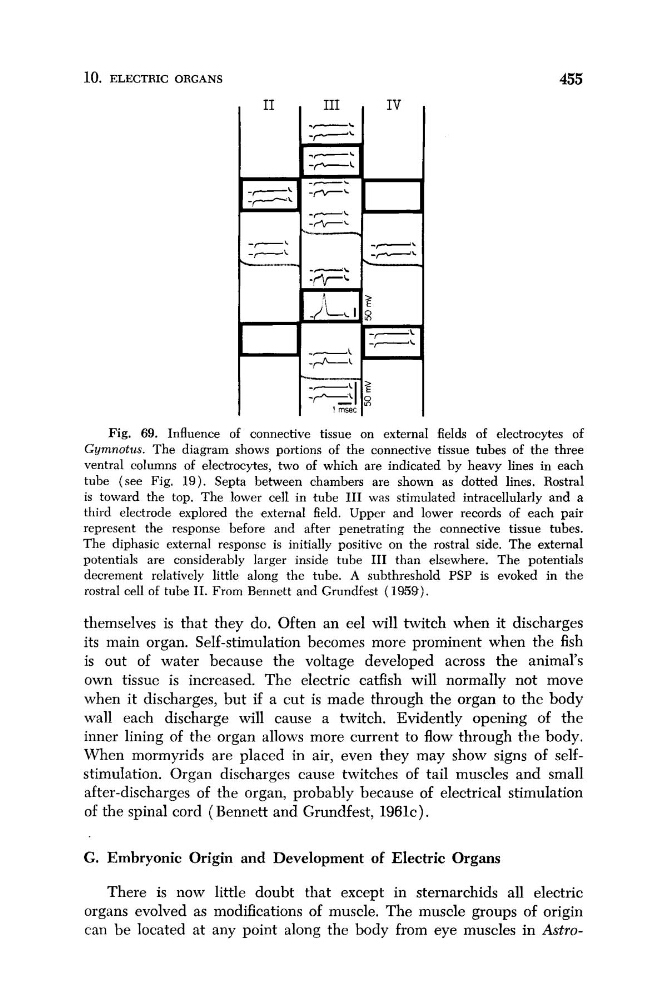
Fig. 69. Influence of connective tissue on external fields of electrocytes of
Gymnotus. The diagram shows portions of the connective tissue tubes of the three
ventral columns of electrocytes, two of which are indicated by heavy lines in each
tube (see Fig. 19). Septa between chambers are shown as dotted lines. Rostra1
is toward the top. The lower cell in tube I11 was stimulated intracellularly and a
third electrode explored the external field. Upper and lower records of each pair
represent the response before and after penetrating the connective tissue tubes.
The diphasic external response is initially positive on the rostral side. The external
potentials are considerably larger inside tube I11 than elsewhere. The potentials
decrement relatively little along the tube. A subthreshold PSP is evoked in the
rostral cell of tube 11. From Bennett and Grundfest (1959).
themselves is that they do. Often an eel will twitch when it discharges
its main organ. Self-stimulation becomes more prominent when the fish
is out of water because the voltage developed across the animal's
own tissue is increased. The electric catfish will normally not move
when it discharges, but if a cut is made through the organ to the body
wall each discharge will cause a twitch. Evidently opening of the
inner lining of the organ allows more current to flow through the body.
When mormyrids are placed in air, even they may show signs of selfstimulation. Organ discharges cause twitches of tail muscles and small
after-discharges of the organ, probably because of electrical stimulation
of the spinal cord (Bennett and Grundfest, 1 9 6 1 ~ ) .
G. Embryonic Origin and Development of Electric Organs
There is now little doubt that except in sternarchids all electric
organs evolved as modifications of muscle. The muscle groups of origin
can be located at any point along the body from eye muscles in Astro-
455
Fig. 69. Influence of connective tissue on external fields of electrocytes of
Gymnotus. The diagram shows portions of the connective tissue tubes of the three
ventral columns of electrocytes, two of which are indicated by heavy lines in each
tube (see Fig. 19). Septa between chambers are shown as dotted lines. Rostra1
is toward the top. The lower cell in tube I11 was stimulated intracellularly and a
third electrode explored the external field. Upper and lower records of each pair
represent the response before and after penetrating the connective tissue tubes.
The diphasic external response is initially positive on the rostral side. The external
potentials are considerably larger inside tube I11 than elsewhere. The potentials
decrement relatively little along the tube. A subthreshold PSP is evoked in the
rostral cell of tube 11. From Bennett and Grundfest (1959).
themselves is that they do. Often an eel will twitch when it discharges
its main organ. Self-stimulation becomes more prominent when the fish
is out of water because the voltage developed across the animal's
own tissue is increased. The electric catfish will normally not move
when it discharges, but if a cut is made through the organ to the body
wall each discharge will cause a twitch. Evidently opening of the
inner lining of the organ allows more current to flow through the body.
When mormyrids are placed in air, even they may show signs of selfstimulation. Organ discharges cause twitches of tail muscles and small
after-discharges of the organ, probably because of electrical stimulation
of the spinal cord (Bennett and Grundfest, 1 9 6 1 ~ ) .
G. Embryonic Origin and Development of Electric Organs
There is now little doubt that except in sternarchids all electric
organs evolved as modifications of muscle. The muscle groups of origin
can be located at any point along the body from eye muscles in Astro-