

10. ELECTRIC ORGANS
407
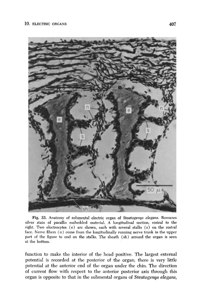
Fig. 33. Anatomy of submental electric organ of Steatogenys elegans. Romanes
silver stain of paraffin embedded material. A longitudinal section, rostral to the
right. Two electrocytes ( e ) are shown, each with several stalks ( s ) on the rostral
face. Nerve fibers ( n ) come from the longitudinally running nerve trunk in the upper
part of the figure to end on the stalks. The sheath ( s h ) around the organ is seen
at the bottom.
function to make the interior of the head positive. The largest external
potential is recorded at the posterior of the organ; there is very little
potential at the anterior end of the organ under the chin. The direction
of current flow with respect to the anterior posterior axis through this
organ is opposite to that in the submental organs of Steatogenys elegans,
407
Fig. 33. Anatomy of submental electric organ of Steatogenys elegans. Romanes
silver stain of paraffin embedded material. A longitudinal section, rostral to the
right. Two electrocytes ( e ) are shown, each with several stalks ( s ) on the rostral
face. Nerve fibers ( n ) come from the longitudinally running nerve trunk in the upper
part of the figure to end on the stalks. The sheath ( s h ) around the organ is seen
at the bottom.
function to make the interior of the head positive. The largest external
potential is recorded at the posterior of the organ; there is very little
potential at the anterior end of the organ under the chin. The direction
of current flow with respect to the anterior posterior axis through this
organ is opposite to that in the submental organs of Steatogenys elegans,