

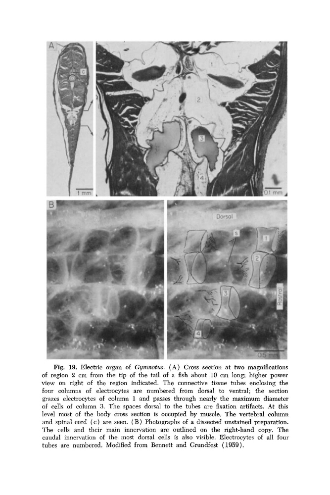
Fig. 19. Electric organ of Gymnotus. (A) Cross section at two magnifications
of region 2 cm from the tip of the tail of a fish about 10 cm long; higher power
view on right of the region indicated. The connective tissue tubes enclosing the
four columns of electrocytes are numbered from dorsal to ventral; the section
grazes electrocytes of column 1 and passes through nearly the maximum diameter
of cells of column 3. The spaces dorsal to the tubes are fixation artifacts. At this
level most of the body cross section is occupied by muscle. The vertebral column
and spinal cord ( c ) are seen. ( B ) Photographs of a dissected unstained preparation.
The cells and their main innervation are outlined on the right-hand copy, The
caudal innervation of the most dorsal cells is also visible. Electrocytes of all four
tubes are numbered. Modified from Bennett and Grundfest (1959).
of region 2 cm from the tip of the tail of a fish about 10 cm long; higher power
view on right of the region indicated. The connective tissue tubes enclosing the
four columns of electrocytes are numbered from dorsal to ventral; the section
grazes electrocytes of column 1 and passes through nearly the maximum diameter
of cells of column 3. The spaces dorsal to the tubes are fixation artifacts. At this
level most of the body cross section is occupied by muscle. The vertebral column
and spinal cord ( c ) are seen. ( B ) Photographs of a dissected unstained preparation.
The cells and their main innervation are outlined on the right-hand copy, The
caudal innervation of the most dorsal cells is also visible. Electrocytes of all four
tubes are numbered. Modified from Bennett and Grundfest (1959).