

A
Rostra1
Caudal
Spinal cord
I Swim. bladder
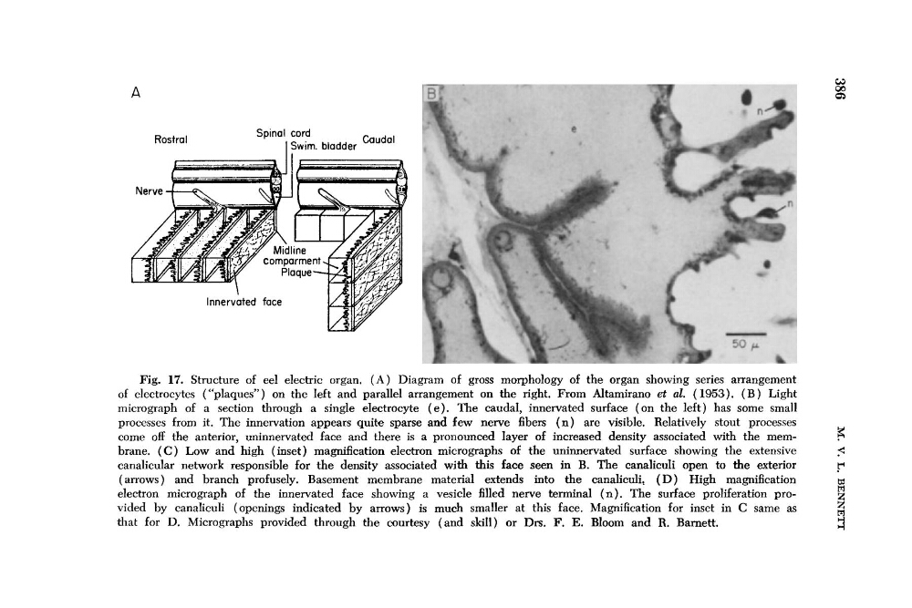
Fig. 17. Structure of eel electric organ, ( A ) Diagram of gross morphology of the organ showing series arrangement
of electrocytes (“plaques”) on the left and parallel arrangement on the right. From Altamirano d al. (1953). ( B ) Light
micrograph of a section through a single electrocyte ( e ) . The caudal, innervated surface (on the left) has some small
processes from it. The innervation appears quite sparse and few nerve fibers ( n ) are visible. Relatively stout processes
come off the anterior, uninnervated face and there is a pronounced layer of increased density associated with the mem5
brane. ( C ) Low and high (inset) magnification electron micrographs of the uninnervated surface showing the extensive
5
canalicular network responsible for the density associated with this face seen in B. The canaliculi open to the exterior
r
(arrows) and branch profusely. Basement membrane material extends into the canaliculi, ( D ) High magnification
m
electron micrograph of the innervated face showing a vesicle filled nerve terminal ( n ) . The surface proliferation provided by canaliculi (openings indicated by arrows) is much smaller at this face. Magnification for inset in C same as
that for D. Micrographs provided through the courtesy (and skill) or Drs. F. E. Bloom and R. Barnett.
H
r-1
Rostra1
Caudal
Spinal cord
I Swim. bladder
Fig. 17. Structure of eel electric organ, ( A ) Diagram of gross morphology of the organ showing series arrangement
of electrocytes (“plaques”) on the left and parallel arrangement on the right. From Altamirano d al. (1953). ( B ) Light
micrograph of a section through a single electrocyte ( e ) . The caudal, innervated surface (on the left) has some small
processes from it. The innervation appears quite sparse and few nerve fibers ( n ) are visible. Relatively stout processes
come off the anterior, uninnervated face and there is a pronounced layer of increased density associated with the mem5
brane. ( C ) Low and high (inset) magnification electron micrographs of the uninnervated surface showing the extensive
5
canalicular network responsible for the density associated with this face seen in B. The canaliculi open to the exterior
r
(arrows) and branch profusely. Basement membrane material extends into the canaliculi, ( D ) High magnification
m
electron micrograph of the innervated face showing a vesicle filled nerve terminal ( n ) . The surface proliferation provided by canaliculi (openings indicated by arrows) is much smaller at this face. Magnification for inset in C same as
that for D. Micrographs provided through the courtesy (and skill) or Drs. F. E. Bloom and R. Barnett.
H
r-1