

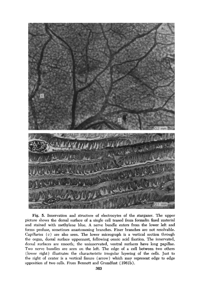
Fig. 5. Innervation and structure of electrocytes of the stargazer. The upper
picture shows the dorsal surface of a single cell teased from formalin fixed material
and stained with methylene blue. A nerve bundle enters from the lower left and
forms profuse, sometimes anastomosing branches. Finer branches are not resolvable.
Capillaries ( c ) are also seen. The lower micrograph is a vertical section through
the organ, dorsal surface uppermost, following osmic acid fixation. The innervated,
dorsal surfaces are smooth; the uninnervated, ventral surfaces have long papillae.
Two nerve bundles are seen on the left. The edge of a cell between two others
(lower right) illustrates the characteristic irregular layering of the cells. Just to
the right of center is a vertical fissure (arrow) which may represent edge to edge
apposition of two cells. From Bennett and Grundfest (196lb).
363
picture shows the dorsal surface of a single cell teased from formalin fixed material
and stained with methylene blue. A nerve bundle enters from the lower left and
forms profuse, sometimes anastomosing branches. Finer branches are not resolvable.
Capillaries ( c ) are also seen. The lower micrograph is a vertical section through
the organ, dorsal surface uppermost, following osmic acid fixation. The innervated,
dorsal surfaces are smooth; the uninnervated, ventral surfaces have long papillae.
Two nerve bundles are seen on the left. The edge of a cell between two others
(lower right) illustrates the characteristic irregular layering of the cells. Just to
the right of center is a vertical fissure (arrow) which may represent edge to edge
apposition of two cells. From Bennett and Grundfest (196lb).
363