

332
J. DIAMOND
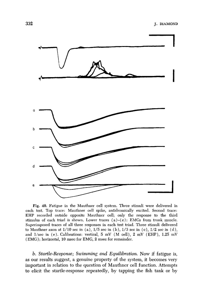
Fig. 40. Fatigue in the Mauthner cell system. Three stimuli were delivered in
each test. Top trace: Mauthner cell spike, antidromically excited. Second trace:
EHP recorded outside opposite Mauthner cell; only the response to the third
stimulus of each triad is shown. Lower traces ( a ) - ( e ) : EMGs from trunk muscle.
Superimposed traces of all three responses in each test triad. Three stimuli delivered
to Mauthner axon at 1/10 sec in ( a ) , 1/5 sec in ( b ) , 1/3 sec in ( c ) , 1/2 sec in ( d ) ,
and l/sec in ( e ) . Calibrations: vertical, 5 mV ( M cell), 2 mV (EHP), 1.25 mV
(EMG); horizontal, 10 m e c for EMG, 2 msec for remainder.
b. Startle-Rc!sponse; Swimming and Equilibration. Now if fatigue is,
as our results suggest, a genuine property of the system, it becomes very
important in relation to the question of Mauthner cell function. Attempts
to elicit the startle-response repeatedly, by tapping the fish tank or by
J. DIAMOND
Fig. 40. Fatigue in the Mauthner cell system. Three stimuli were delivered in
each test. Top trace: Mauthner cell spike, antidromically excited. Second trace:
EHP recorded outside opposite Mauthner cell; only the response to the third
stimulus of each triad is shown. Lower traces ( a ) - ( e ) : EMGs from trunk muscle.
Superimposed traces of all three responses in each test triad. Three stimuli delivered
to Mauthner axon at 1/10 sec in ( a ) , 1/5 sec in ( b ) , 1/3 sec in ( c ) , 1/2 sec in ( d ) ,
and l/sec in ( e ) . Calibrations: vertical, 5 mV ( M cell), 2 mV (EHP), 1.25 mV
(EMG); horizontal, 10 m e c for EMG, 2 msec for remainder.
b. Startle-Rc!sponse; Swimming and Equilibration. Now if fatigue is,
as our results suggest, a genuine property of the system, it becomes very
important in relation to the question of Mauthner cell function. Attempts
to elicit the startle-response repeatedly, by tapping the fish tank or by