

9. THE MAUTHNER CELL
I 2
285
J
A
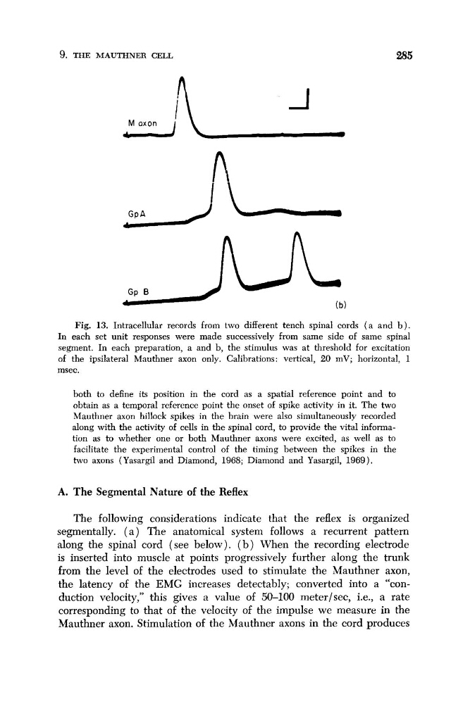
Fig. 13. Intracellular records from two different tench spinal cords ( a and b ) .
In each set unit responses were made successively from same side of same spinal
segment. In each preparation, a and b, the stimulus was at threshold for excitation
of the ipsilateral Mauthner axon only. Calibrations: vertical, 20 mV; horizontal, 1
msec.
both to define its position in the cord as a spatial reference point and to
obtain as a temporal reference point the onset of spike activity in it. The two
Mauthner axon hillock spikes in the brain were also simultaneously recorded
along with the activity of cells in the spinal cord, to provide the vital information as to whether one or both Mauthner axons were excited, as well as to
facilitate the experimental control of the timing between the spikes in the
two axons (Yasargil and Diamond, 1968; Diamond and Yasargil, 1969).
A. The Segmental Nature of the Reflex
The following considerations indicate that the reflex is organized
segmentally. ( a ) The anatomical system follows a recurrent pattern
along the spinal cord (see below). ( b ) When the recording electrode
is inserted into muscle at points progressively further along the trunk
from the level of the electrodes used to stimulate the Mauthner axon,
the latency of the EMG increases detectably; converted into a “conduction velocity,” this gives a value of 50-100 meterlsec, i.e., a rate
corresponding to that of the velocity o€ the impulse we measure in the
Mauthner axon. Stimulation of the Mauthner axons in the cord produces
I 2
285
J
A
Fig. 13. Intracellular records from two different tench spinal cords ( a and b ) .
In each set unit responses were made successively from same side of same spinal
segment. In each preparation, a and b, the stimulus was at threshold for excitation
of the ipsilateral Mauthner axon only. Calibrations: vertical, 20 mV; horizontal, 1
msec.
both to define its position in the cord as a spatial reference point and to
obtain as a temporal reference point the onset of spike activity in it. The two
Mauthner axon hillock spikes in the brain were also simultaneously recorded
along with the activity of cells in the spinal cord, to provide the vital information as to whether one or both Mauthner axons were excited, as well as to
facilitate the experimental control of the timing between the spikes in the
two axons (Yasargil and Diamond, 1968; Diamond and Yasargil, 1969).
A. The Segmental Nature of the Reflex
The following considerations indicate that the reflex is organized
segmentally. ( a ) The anatomical system follows a recurrent pattern
along the spinal cord (see below). ( b ) When the recording electrode
is inserted into muscle at points progressively further along the trunk
from the level of the electrodes used to stimulate the Mauthner axon,
the latency of the EMG increases detectably; converted into a “conduction velocity,” this gives a value of 50-100 meterlsec, i.e., a rate
corresponding to that of the velocity o€ the impulse we measure in the
Mauthner axon. Stimulation of the Mauthner axons in the cord produces