

14
F. W. MUNZ
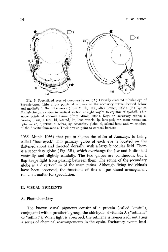
Fig. 5. Specialized eyes of deep-sea fishes. ( A ) Dorsally directed tubular eye of
Scopelarchus. Thin arrow points at a piece of the accessory retina located below
and medially to the optic nerve (from Munk, 1966, after Brauer, 1908). ( B ) Eye of
Bathylychnops as seen in vertical section at right angles to equator of eyeball. Thin
arrow points at choroid fissure (from Munk, 1966). Key: ar, accessory retina; c,
cornea; i, iris; 1, lens; Id, laterad; lm, lens muscle; Ip, lens-pad; mr, main retina; on,
optic nerve; r, retina; s, sclera; sg, secondary globe; sl, scleral lens; and w, window
of the diverticulnm-retina. Thick arrows point to corneal borders.
1965; Munk, 1966) that put to shame the claim of AnabZeps to being
called “four-eyed.” The primary globe of each eye is located on the
flattened snout and directed dorsally, with a large binocular field. There
is a secondary globe (Fig. 5B), which overhangs the jaw and is directed
ventrally and slightly caudally. The two globes are continuous, but a
flap keeps light from passing between them. The retina of the secondary
globe is a diverticulum of the main retina. Although living individuals
have been observed, the functions of this unique visual arrangement
remain a matter for speculation.
11. VISUAL PIGMENTS
A. Photochemistry
The known visual pigments consist of a protein (called “opsin”),
conjugated with a prosthetic group, the aldehyde of vitamin A (“retinene”
or “retinal”). When light is absorbed, the retinene is isomerized, initiating
a series of chemical rearrangements in the opsin. Excitatory events lead-
F. W. MUNZ
Fig. 5. Specialized eyes of deep-sea fishes. ( A ) Dorsally directed tubular eye of
Scopelarchus. Thin arrow points at a piece of the accessory retina located below
and medially to the optic nerve (from Munk, 1966, after Brauer, 1908). ( B ) Eye of
Bathylychnops as seen in vertical section at right angles to equator of eyeball. Thin
arrow points at choroid fissure (from Munk, 1966). Key: ar, accessory retina; c,
cornea; i, iris; 1, lens; Id, laterad; lm, lens muscle; Ip, lens-pad; mr, main retina; on,
optic nerve; r, retina; s, sclera; sg, secondary globe; sl, scleral lens; and w, window
of the diverticulnm-retina. Thick arrows point to corneal borders.
1965; Munk, 1966) that put to shame the claim of AnabZeps to being
called “four-eyed.” The primary globe of each eye is located on the
flattened snout and directed dorsally, with a large binocular field. There
is a secondary globe (Fig. 5B), which overhangs the jaw and is directed
ventrally and slightly caudally. The two globes are continuous, but a
flap keeps light from passing between them. The retina of the secondary
globe is a diverticulum of the main retina. Although living individuals
have been observed, the functions of this unique visual arrangement
remain a matter for speculation.
11. VISUAL PIGMENTS
A. Photochemistry
The known visual pigments consist of a protein (called “opsin”),
conjugated with a prosthetic group, the aldehyde of vitamin A (“retinene”
or “retinal”). When light is absorbed, the retinene is isomerized, initiating
a series of chemical rearrangements in the opsin. Excitatory events lead-