

9. THE MAUTHNER CELL
269
200 / A.
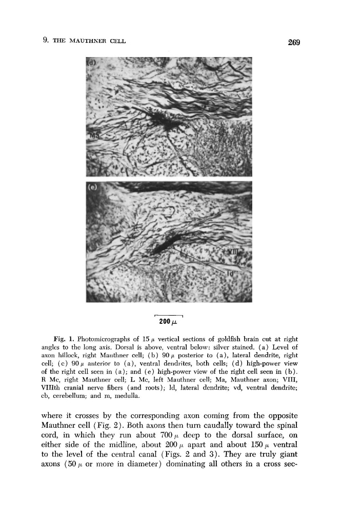
Fig. 1. Photomicrographs of 1 5 p vertical sections of goldfish brain cut at right
angles to the long axis. Dorsal is above, ventral below: silver stained. ( a ) Level of
axon hillock, right Mauthner cell; ( b ) 9 O p posterior to ( a ) , lateral dendrite, right
cell; ( c ) 90 p anterior to ( a ) , ventral dendrites, both cells; ( d ) high-power view
of the right cell seen in ( a ) ; and ( e ) high-power view of the right cell seen in ( b ) .
R Mc, right Mauthner cell; L Mc, left Mauthner cell; Ma, Mauthner axon; VIII,
VIIIth cranial nerve fibers (and roots); Id, lateral dendrite; vd, ventral dendrite;
cb, cerebellum; and m, medulla.
where it crosses by the corresponding axon coming from the opposite
Mauthner cell (Fig. 2 ) . Both axons then turn caudally toward the spinal
cord, in which they run about 700p deep to the dorsal surface, on
either side of the midline, about 200p apart and about 1 5 0 p ventral
to the level of the central canal (Figs. 2 and 3 ) . They are truly giant
axons (50 ,LL or more in diameter) dominating all others in a cross sec-
269
200 / A.
Fig. 1. Photomicrographs of 1 5 p vertical sections of goldfish brain cut at right
angles to the long axis. Dorsal is above, ventral below: silver stained. ( a ) Level of
axon hillock, right Mauthner cell; ( b ) 9 O p posterior to ( a ) , lateral dendrite, right
cell; ( c ) 90 p anterior to ( a ) , ventral dendrites, both cells; ( d ) high-power view
of the right cell seen in ( a ) ; and ( e ) high-power view of the right cell seen in ( b ) .
R Mc, right Mauthner cell; L Mc, left Mauthner cell; Ma, Mauthner axon; VIII,
VIIIth cranial nerve fibers (and roots); Id, lateral dendrite; vd, ventral dendrite;
cb, cerebellum; and m, medulla.
where it crosses by the corresponding axon coming from the opposite
Mauthner cell (Fig. 2 ) . Both axons then turn caudally toward the spinal
cord, in which they run about 700p deep to the dorsal surface, on
either side of the midline, about 200p apart and about 1 5 0 p ventral
to the level of the central canal (Figs. 2 and 3 ) . They are truly giant
axons (50 ,LL or more in diameter) dominating all others in a cross sec-