

250
h FLOCK
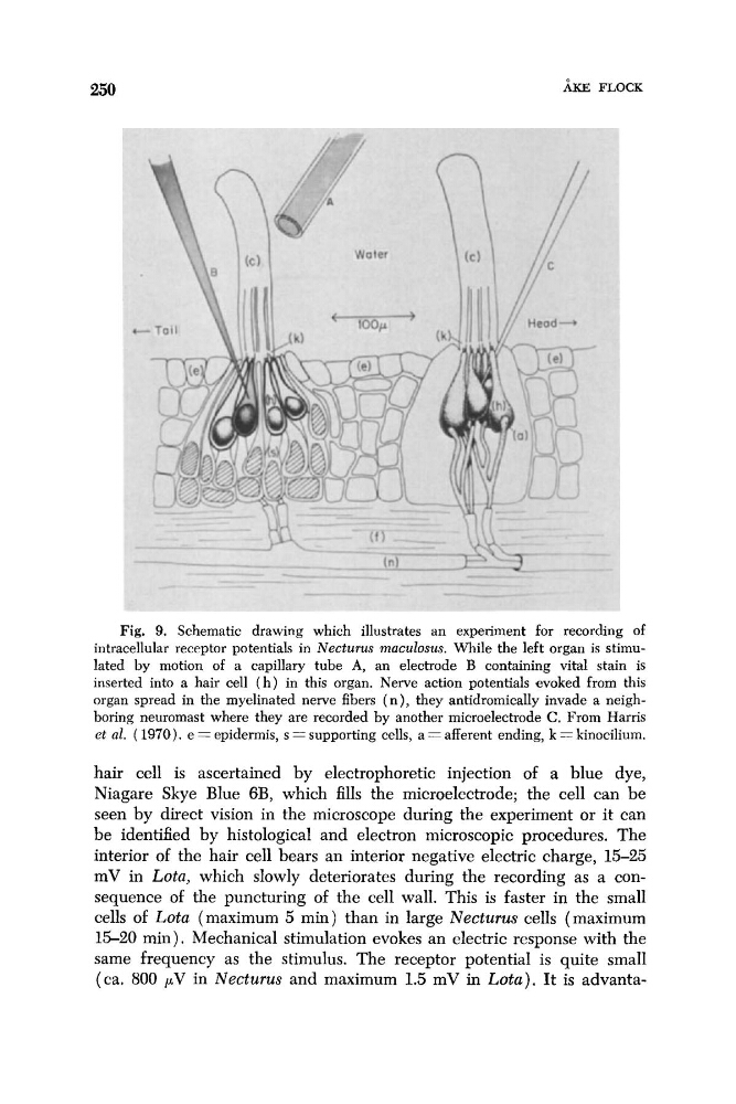
Fig. 9. Schematic drawing which illustrates an experiment for recording of
intracellular receptor potentials in Necturus rnaculosus. While the left organ is stimulated by motion of a capillary tube A, an electrode B containing vital stain is
inserted into a hair cell ( h ) in this organ. Nerve action potentials evoked from this
organ spread in the myelinated nerve fibers ( n ) , they antidromically invade a neighboring neuromast where they are recorded by another microelectrode C. From Harris
et al. ( 1970). e = epidermis, s = supporting cells, a = afferent ending, k = kinocilium.
hair cell is ascertained by electrophoretic injection of a blue dye,
Niagare Skye Blue 6B, which fills the microelectrode; the cell can be
seen by direct vision in the microscope during the experiment or it can
be identified by histological and electron microscopic procedures. The
interior of the hair cell bears an interior negative electric charge, 15-25
mV in Lota, which slowly deteriorates during the recording as a consequence of the puncturing of the cell wall. This is faster in the small
cells of Lota (maximum 5 min) than in large Necturus cells (maximum
1 5 2 0 min) . Mechanical stimulation evokes an electric response with the
same frequency as the stimulus. The receptor potential is quite small
(ca. 800 pV in Necturus and maximum 1.5 mV in Lota). It is advanta-
h FLOCK
Fig. 9. Schematic drawing which illustrates an experiment for recording of
intracellular receptor potentials in Necturus rnaculosus. While the left organ is stimulated by motion of a capillary tube A, an electrode B containing vital stain is
inserted into a hair cell ( h ) in this organ. Nerve action potentials evoked from this
organ spread in the myelinated nerve fibers ( n ) , they antidromically invade a neighboring neuromast where they are recorded by another microelectrode C. From Harris
et al. ( 1970). e = epidermis, s = supporting cells, a = afferent ending, k = kinocilium.
hair cell is ascertained by electrophoretic injection of a blue dye,
Niagare Skye Blue 6B, which fills the microelectrode; the cell can be
seen by direct vision in the microscope during the experiment or it can
be identified by histological and electron microscopic procedures. The
interior of the hair cell bears an interior negative electric charge, 15-25
mV in Lota, which slowly deteriorates during the recording as a consequence of the puncturing of the cell wall. This is faster in the small
cells of Lota (maximum 5 min) than in large Necturus cells (maximum
1 5 2 0 min) . Mechanical stimulation evokes an electric response with the
same frequency as the stimulus. The receptor potential is quite small
(ca. 800 pV in Necturus and maximum 1.5 mV in Lota). It is advanta-