

224
0. LOWENSTEIN
Dorsal
Anterior horizontal rnacula
Median
Lateral
Posterior
Ventral
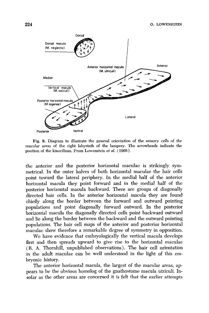
Fig. 9. Diagram to illustrate the general orientation of the sensory cells of the
macular areas of the right labyrinth of the lamprey. The arrowheads indicate the
position of the kinocilium. From Lowenstein et al. ( 1968).
the anterior and the posterior horizontal maculae is strikingly symmetrical. In the outer halves of both horizontal maculae the hair cells
point toward the lateral periphery. In the medial half of the anterior
horizontal macula they point forward and in the medial half of the
posterior horizontal macula backward. There are groups of diagonally
directed hair cells. In the anterior horizontal macula they are found
chiefly along the border between the forward and outward pointing
populations and point diagonally forward outward. In the posterior
horizontal macula the diagonally directed cells point backward outward
and lie along the border between the backward and the outward pointing
populations. The hair cell maps of the anterior and posterior horizontal
maculae show therefore a remarkable degree of symmetry in opposition.
We have evidence that embryologically the vertical macula develops
first and then spreads upward to give rise to the horizontal maculae
( R. A. Thornhill, unpublished observations). The hair cell orientation
in the adult maculae can be well understood in the light of this embryonic history.
The anterior horizontal macula, the largest of the macular areas, appears to be the obvious homolog of the gnathostome macula utriculi. Insofar as the other areas are concerned it is felt that the earlier attempts
0. LOWENSTEIN
Dorsal
Anterior horizontal rnacula
Median
Lateral
Posterior
Ventral
Fig. 9. Diagram to illustrate the general orientation of the sensory cells of the
macular areas of the right labyrinth of the lamprey. The arrowheads indicate the
position of the kinocilium. From Lowenstein et al. ( 1968).
the anterior and the posterior horizontal maculae is strikingly symmetrical. In the outer halves of both horizontal maculae the hair cells
point toward the lateral periphery. In the medial half of the anterior
horizontal macula they point forward and in the medial half of the
posterior horizontal macula backward. There are groups of diagonally
directed hair cells. In the anterior horizontal macula they are found
chiefly along the border between the forward and outward pointing
populations and point diagonally forward outward. In the posterior
horizontal macula the diagonally directed cells point backward outward
and lie along the border between the backward and the outward pointing
populations. The hair cell maps of the anterior and posterior horizontal
maculae show therefore a remarkable degree of symmetry in opposition.
We have evidence that embryologically the vertical macula develops
first and then spreads upward to give rise to the horizontal maculae
( R. A. Thornhill, unpublished observations). The hair cell orientation
in the adult maculae can be well understood in the light of this embryonic history.
The anterior horizontal macula, the largest of the macular areas, appears to be the obvious homolog of the gnathostome macula utriculi. Insofar as the other areas are concerned it is felt that the earlier attempts