

0. LOWENSTEIN
208
A
PC
cs
A
C
L
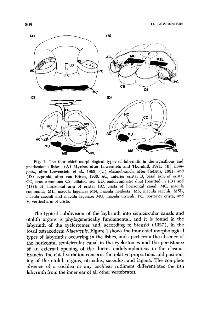
Fig. 1. The four chief morphological types of labyrinth in the agnathous and
gnathostome fishes. ( A ) Myxine, after Lowenstein and Thornhill, 1971; ( B ) Lampetru, after Lowenstein et al., 1968; ( C ) elasmobranch, after Retzius, 1881; and
( D ) cyprinid, after von Frisch, 1936. AC, anterior crista; B, basal arm of crista;
CC, crus commune; CS, ciliated sac; ED, endolymphatic duct [omitted in ( B ) and
( D ) ] ; H, horizontal arm of crista; HC, crista of horizontal canal; MC, macula
communis; ML, macula lagenae; MN, macula neglecta; MS, macula sacculi; MSL,
macula sacculi and macula lagenae; MU, macula utriculi; PC, posterior crista; and
V, vertical arm of crista.
The typical subdivision of the laybrinth into semicircular canals and
otolith organs is phylogenetically fundamental, and it is found in the
labyrinth of the cyclostomes and, according to Stensi6 (1927), in the
fossil ostracoderm Kiaermpis. Figure 1 shows the four chief morphological
types of labyrinths occurring in the fishes, and apart from the absence of
the horizontal semicircular canal in the cyclostomes and the persistence
of an external opening of the ductus endolymphaticus in the elasmobranchs, the chief variation concerns the relative proportions and positioning of the otolith organs, utriculus, sacculus, and lagena. The complete
absence of a cochlea or any cochlear rudiment differentiates the fish
labyrinth from the inner ear of all other vertebrates.
208
A
PC
cs
A
C
L
Fig. 1. The four chief morphological types of labyrinth in the agnathous and
gnathostome fishes. ( A ) Myxine, after Lowenstein and Thornhill, 1971; ( B ) Lampetru, after Lowenstein et al., 1968; ( C ) elasmobranch, after Retzius, 1881; and
( D ) cyprinid, after von Frisch, 1936. AC, anterior crista; B, basal arm of crista;
CC, crus commune; CS, ciliated sac; ED, endolymphatic duct [omitted in ( B ) and
( D ) ] ; H, horizontal arm of crista; HC, crista of horizontal canal; MC, macula
communis; ML, macula lagenae; MN, macula neglecta; MS, macula sacculi; MSL,
macula sacculi and macula lagenae; MU, macula utriculi; PC, posterior crista; and
V, vertical arm of crista.
The typical subdivision of the laybrinth into semicircular canals and
otolith organs is phylogenetically fundamental, and it is found in the
labyrinth of the cyclostomes and, according to Stensi6 (1927), in the
fossil ostracoderm Kiaermpis. Figure 1 shows the four chief morphological
types of labyrinths occurring in the fishes, and apart from the absence of
the horizontal semicircular canal in the cyclostomes and the persistence
of an external opening of the ductus endolymphaticus in the elasmobranchs, the chief variation concerns the relative proportions and positioning of the otolith organs, utriculus, sacculus, and lagena. The complete
absence of a cochlea or any cochlear rudiment differentiates the fish
labyrinth from the inner ear of all other vertebrates.