

88
TOSHTAKI J. HAM
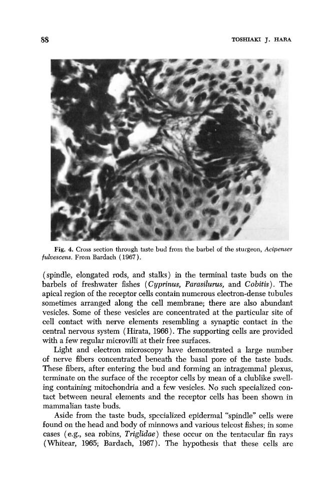
Fig. 4. Cross section through taste bud from the barbel of the sturgeon, Acipeiiser
fulvescens. From Bardach (1967).
(spindle, elongated rods, and stalks) in the terminal taste buds on the
barbels of freshwater fishes ( Cyprinus, Purusilurus, and Cobitis). The
apical region of the receptor cells contain numerous electron-dense tubules
sometimes arranged along the cell membrane; there are also abundant
vesicles. Some of these vesicles are concentrated at the particular site of
cell contact with nerve elements resembling a synaptic contact in the
central nervous system ( Hirata, 1966). The supporting cells are provided
with a few regular microvilli at their free surfaces.
Light and electron microscopy have demonstrated a large number
of nerve fibers concentrated beneath the basal pore of the taste buds.
These fibers, after entering the bud and forming an intragemmal plexus,
terminate on the surface of the receptor cells by mean of a clublike swelling containing mitochondria and a few vesicles. No such specialized contact between neural elements and the receptor cells has been shown in
mammalian taste buds.
Aside from the taste buds, specialized epidermal “spindle” cells were
found on the head and body of minnows and various teleost fishes; in some
cases (e.g., sea robins, Triglidue) these occur on the tentacular fin rays
(Whitear, 1965; Bardach, 1967). The hypothesis that these cells are
TOSHTAKI J. HAM
Fig. 4. Cross section through taste bud from the barbel of the sturgeon, Acipeiiser
fulvescens. From Bardach (1967).
(spindle, elongated rods, and stalks) in the terminal taste buds on the
barbels of freshwater fishes ( Cyprinus, Purusilurus, and Cobitis). The
apical region of the receptor cells contain numerous electron-dense tubules
sometimes arranged along the cell membrane; there are also abundant
vesicles. Some of these vesicles are concentrated at the particular site of
cell contact with nerve elements resembling a synaptic contact in the
central nervous system ( Hirata, 1966). The supporting cells are provided
with a few regular microvilli at their free surfaces.
Light and electron microscopy have demonstrated a large number
of nerve fibers concentrated beneath the basal pore of the taste buds.
These fibers, after entering the bud and forming an intragemmal plexus,
terminate on the surface of the receptor cells by mean of a clublike swelling containing mitochondria and a few vesicles. No such specialized contact between neural elements and the receptor cells has been shown in
mammalian taste buds.
Aside from the taste buds, specialized epidermal “spindle” cells were
found on the head and body of minnows and various teleost fishes; in some
cases (e.g., sea robins, Triglidue) these occur on the tentacular fin rays
(Whitear, 1965; Bardach, 1967). The hypothesis that these cells are