

8. UROPHYSIS AND CAUDAL NEUROSECRETORY SYSTEM
409
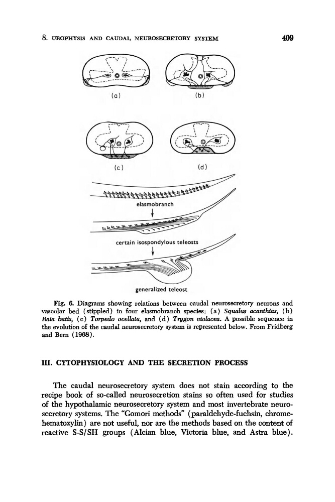
Fig. 6. Diagrams showing relations between caudal neurosecretory neurons and
vascular bed (stippled) in four elasmobranch species: ( a ) Sqwlus acanthias, (b)
Raia batis, ( c ) Torpedo ocelhtu, and ( d ) Tvgon violacea. A possible sequence in
the evolution of the caudal neurosecretory system is represented below. From Fridberg
and Bern (1988).
III. CYTOPHYSIOLOGY AND THE SECRETION PROCESS
The caudal neurosecretory system does not stain according to the
recipe book of so-called neurosecretion stains so often used for studies
of the hypothalamic neurosecretory system and most invertebrate neurosecretory systems. The “Gomori methods” ( paraldehyde-fuchsin, chromehematoxylin) are not useful, nor are the methods based on the content of
reactive S-S/SH groups (Alcian blue, Victoria blue, and Astra blue).
409
Fig. 6. Diagrams showing relations between caudal neurosecretory neurons and
vascular bed (stippled) in four elasmobranch species: ( a ) Sqwlus acanthias, (b)
Raia batis, ( c ) Torpedo ocelhtu, and ( d ) Tvgon violacea. A possible sequence in
the evolution of the caudal neurosecretory system is represented below. From Fridberg
and Bern (1988).
III. CYTOPHYSIOLOGY AND THE SECRETION PROCESS
The caudal neurosecretory system does not stain according to the
recipe book of so-called neurosecretion stains so often used for studies
of the hypothalamic neurosecretory system and most invertebrate neurosecretory systems. The “Gomori methods” ( paraldehyde-fuchsin, chromehematoxylin) are not useful, nor are the methods based on the content of
reactive S-S/SH groups (Alcian blue, Victoria blue, and Astra blue).