

404
HOWARD A. BERN
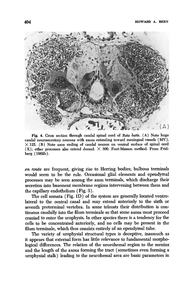
Fig. 4. Cross section through caudal spinal cord of Roiu both. ( A ) Note huge
caudal neurosecretory neurons with s o n s extending toward meningeal vessels (MV).
X 125. ( B ) Note axon ending of caudal neuron on ventral surface of spinal cord
( X ) ; other processes also extend dorsad. X 300. Foot-Masson method. From Fridberg (1962b).
en route are frequent, giving rise to Herring bodies; bulbous terminals
would seem to be the rule. Occasional glial elements and ependymal
processes may be seen among the axon terminals, which discharge their
secretion into basement membrane regions intervening between them and
the capillary endothelium (Fig. 3).
The cell somata (Fig. 1D) of the system are generally located ventrolateral to the central canal and may extend anteriorly to the sixth or
seventh preterminal vertebra. In some teleosts their distribution is continuous caudally into the filum terminale so that some axons must proceed
craniad to enter the urophysis. In other species there is a tendency for the
cells to be concentrated anteriorly, and no cells may be present in the
6lum terminale, which thus consists entirely of an ependymal tube.
The variety of urophysial structural types is deceptive, inasmuch as
it appears that external form has little relevance to fundamental morphological differences. The relation of the neurohemal region to the meninx
and the length of the axons forming the tract (sometimes even forming a
urophysial stalk) leading to the neurohemal area are basic parameters in
HOWARD A. BERN
Fig. 4. Cross section through caudal spinal cord of Roiu both. ( A ) Note huge
caudal neurosecretory neurons with s o n s extending toward meningeal vessels (MV).
X 125. ( B ) Note axon ending of caudal neuron on ventral surface of spinal cord
( X ) ; other processes also extend dorsad. X 300. Foot-Masson method. From Fridberg (1962b).
en route are frequent, giving rise to Herring bodies; bulbous terminals
would seem to be the rule. Occasional glial elements and ependymal
processes may be seen among the axon terminals, which discharge their
secretion into basement membrane regions intervening between them and
the capillary endothelium (Fig. 3).
The cell somata (Fig. 1D) of the system are generally located ventrolateral to the central canal and may extend anteriorly to the sixth or
seventh preterminal vertebra. In some teleosts their distribution is continuous caudally into the filum terminale so that some axons must proceed
craniad to enter the urophysis. In other species there is a tendency for the
cells to be concentrated anteriorly, and no cells may be present in the
6lum terminale, which thus consists entirely of an ependymal tube.
The variety of urophysial structural types is deceptive, inasmuch as
it appears that external form has little relevance to fundamental morphological differences. The relation of the neurohemal region to the meninx
and the length of the axons forming the tract (sometimes even forming a
urophysial stalk) leading to the neurohemal area are basic parameters in