

7. THE ULTIMOBRANCHIAL GLANDS
381
Thvmus
Elasmobranchs
Teleosts
Thymus
Amphibians
Q$Jip Ultimobronchial
Cumlid
my Pornthyroids Aortic body
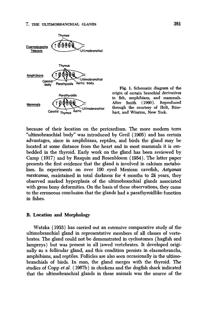
Fig. 1. Schematic diagram of the
origin of certain branchial derivatives
in fish, amphibians, and mammals.
After Smith (1960). Reproduced
through the courtesy of Holt, Rinehart, and Winston, New York.
Parathyroids
imbranchial
Mammals
because of their location on the pericardium. The more modem term
“ultimobranchial body” was introduced by Greil (1905) and has certain
advantages, since in amphibians, reptiles, and birds the gland may be
located at some distance from the heart and in most mammals it is embedded in the thyroid. Early work on the gland has been reviewed by
Camp (1917) and by Rasquin and Rosenbloom (1954). The latter paper
presents the first evidence that the gland is involved in calcium metabolism. In experiments on over 100 eyed Mexican cavefish, Astyam
mexicanus, maintained in total darkness for 4 months to W years, they
observed marked hyperplasia of the ultimobranchial glands associated
with gross bony deformities. On the basis of these observations, they came
to the erroneous conclusion that the glands had a parathyroidlike function
in fishes.
B. Location and Morphology
Watzka (1933) has carried out an extensive comparative study of the
ultimobranchial gland in representative members of all classes of vertebrates. The gland could not be demonstrated in cyclostomes (hagfish and
lampreys) but was present in all jawed vertebrates. It developed originally as a follicular gland, and this condition persists in elasmobranchs,
amphibians, and reptiles. Follicles are also seen occasionally in the ultimobranchials of birds. In man, the gland merges with the thyroid. The
studies of Copp et al. ( 1967b) in chickens and the dogfish shark indicated
that the ultimobranchial glands in these animals was the source of the
381
Thvmus
Elasmobranchs
Teleosts
Thymus
Amphibians
Q$Jip Ultimobronchial
Cumlid
my Pornthyroids Aortic body
Fig. 1. Schematic diagram of the
origin of certain branchial derivatives
in fish, amphibians, and mammals.
After Smith (1960). Reproduced
through the courtesy of Holt, Rinehart, and Winston, New York.
Parathyroids
imbranchial
Mammals
because of their location on the pericardium. The more modem term
“ultimobranchial body” was introduced by Greil (1905) and has certain
advantages, since in amphibians, reptiles, and birds the gland may be
located at some distance from the heart and in most mammals it is embedded in the thyroid. Early work on the gland has been reviewed by
Camp (1917) and by Rasquin and Rosenbloom (1954). The latter paper
presents the first evidence that the gland is involved in calcium metabolism. In experiments on over 100 eyed Mexican cavefish, Astyam
mexicanus, maintained in total darkness for 4 months to W years, they
observed marked hyperplasia of the ultimobranchial glands associated
with gross bony deformities. On the basis of these observations, they came
to the erroneous conclusion that the glands had a parathyroidlike function
in fishes.
B. Location and Morphology
Watzka (1933) has carried out an extensive comparative study of the
ultimobranchial gland in representative members of all classes of vertebrates. The gland could not be demonstrated in cyclostomes (hagfish and
lampreys) but was present in all jawed vertebrates. It developed originally as a follicular gland, and this condition persists in elasmobranchs,
amphibians, and reptiles. Follicles are also seen occasionally in the ultimobranchials of birds. In man, the gland merges with the thyroid. The
studies of Copp et al. ( 1967b) in chickens and the dogfish shark indicated
that the ultimobranchial glands in these animals was the source of the