

J. N. BALL AND BRIDGET I. BAKER
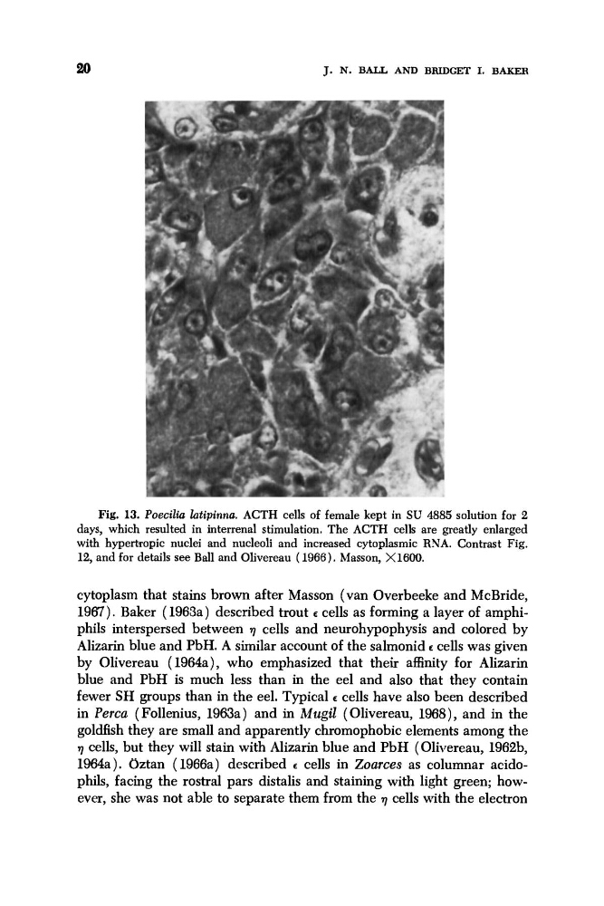
Fig. 13. Poecilia latipinnu. ACTH cells of female kept in SU 4885 solution for 2
days, which resulted in interrenal stimulation. The ACTH cells are greatly enlarged
with hypertropic nuclei and nucleoli and increased cytoplasmic RNA. Contrast Fig.
12, and for details see Ball and Olivereau (1966). Masson, X1600.
cytoplasm that stains brown after Masson (van Overbeeke and McBride,
1967). Baker (1963a) described trout c cells as forming a layer of amphiphils interspersed between 7 cells and neurohypophysis and colored by
Alizarin blue and PbH. A similar account of the salmonid c cells was given
by Olivereau (1964a), who emphasized that their affinity for Alizarin
blue and PbH is much less than in the eel and also that they contain
fewer SH groups than in the eel. Typical c cells have also been described
in Perm (Follenius, 1963a) and in Mugil (Olivereau, 1968), and in the
goldfish they are small and apparently chromophobic elements among the
7 cells, but they will stain with Alizarin blue and PbH (Olivereau, 1962b,
1964a). Oztan (1966a) described L cells in Zoarces as columnar acidophils, facing the rostra1 pars distalis and staining with light green; however, she was not able to separate them from the 7 cells with the electron
Fig. 13. Poecilia latipinnu. ACTH cells of female kept in SU 4885 solution for 2
days, which resulted in interrenal stimulation. The ACTH cells are greatly enlarged
with hypertropic nuclei and nucleoli and increased cytoplasmic RNA. Contrast Fig.
12, and for details see Ball and Olivereau (1966). Masson, X1600.
cytoplasm that stains brown after Masson (van Overbeeke and McBride,
1967). Baker (1963a) described trout c cells as forming a layer of amphiphils interspersed between 7 cells and neurohypophysis and colored by
Alizarin blue and PbH. A similar account of the salmonid c cells was given
by Olivereau (1964a), who emphasized that their affinity for Alizarin
blue and PbH is much less than in the eel and also that they contain
fewer SH groups than in the eel. Typical c cells have also been described
in Perm (Follenius, 1963a) and in Mugil (Olivereau, 1968), and in the
goldfish they are small and apparently chromophobic elements among the
7 cells, but they will stain with Alizarin blue and PbH (Olivereau, 1962b,
1964a). Oztan (1966a) described L cells in Zoarces as columnar acidophils, facing the rostra1 pars distalis and staining with light green; however, she was not able to separate them from the 7 cells with the electron