

178
A. M. PERKS
the tetrapods ( W. H. Sawyer, 1966b). Although the amphibians have not
descended directly from the lungfish, it is likely that they shared a
common crossopterygian ancestor ( Follet and Heller, 1964b), and until
the pituitaries of crossopterygian species become available for study, our
picture of the evolution of the neurohypophysis from fish to tetrapod
must depend on investigations of the lungfish. As early as 1926, de Beer
was struck by the strong resemblance between the lungfish pituitary and
that of the amphibians. Since this time, many workers have made similar
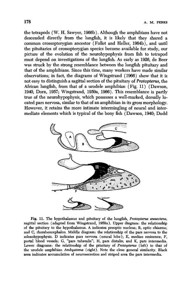
observations; in fact, the diagrams of Wingstrand (1W) show that it is
not easy to distinguish a sagittal section of the pituitary of Protopterus, the
African lungfish, from that of a urodele amphibian (Fig. 11) (Dawson,
1940, Dorn, 1957; Wingstrand, 1959a, 1966). This resemblance is partly
true of the neurohypophysis, which possesses a well-marked, dorsally located pars nervosa, similar to that of an amphibian in its gross morphology.
However, it retains the more intimate intermingling of neural and intermediate elements which is typical of the bony fish (Dawson, 1940; Dodd
Fig. 11. The hypothalamus and pituitary of the lungfish, Protoptern annectens,
sagittal section (adapted from Wingstrand, 1959a). Upper diagram: the relationship
of the pituitary to the hypothalamus. A indicates preoptic nucleus; B, optic chiasma;
and C, rhombencephalon. Middle diagram: the relationship of the pars nervosa to the
adenohypophysis. D indicates pars nervosa (neural lobe); E, median eminence; F,
portal blood vessels; G, “pars tuberalis”; H, pars distalis; and K, pars intermedia.
Lower diagrams: the relationship of the pituitary of Pmtopterus (left) to that of
the urodele amphibian Ambystoma (right). Note the close general similarity. Black
area indicates accumulation of neurosecretion and striped area the pars intermedia.
A. M. PERKS
the tetrapods ( W. H. Sawyer, 1966b). Although the amphibians have not
descended directly from the lungfish, it is likely that they shared a
common crossopterygian ancestor ( Follet and Heller, 1964b), and until
the pituitaries of crossopterygian species become available for study, our
picture of the evolution of the neurohypophysis from fish to tetrapod
must depend on investigations of the lungfish. As early as 1926, de Beer
was struck by the strong resemblance between the lungfish pituitary and
that of the amphibians. Since this time, many workers have made similar
observations; in fact, the diagrams of Wingstrand (1W) show that it is
not easy to distinguish a sagittal section of the pituitary of Protopterus, the
African lungfish, from that of a urodele amphibian (Fig. 11) (Dawson,
1940, Dorn, 1957; Wingstrand, 1959a, 1966). This resemblance is partly
true of the neurohypophysis, which possesses a well-marked, dorsally located pars nervosa, similar to that of an amphibian in its gross morphology.
However, it retains the more intimate intermingling of neural and intermediate elements which is typical of the bony fish (Dawson, 1940; Dodd
Fig. 11. The hypothalamus and pituitary of the lungfish, Protoptern annectens,
sagittal section (adapted from Wingstrand, 1959a). Upper diagram: the relationship
of the pituitary to the hypothalamus. A indicates preoptic nucleus; B, optic chiasma;
and C, rhombencephalon. Middle diagram: the relationship of the pars nervosa to the
adenohypophysis. D indicates pars nervosa (neural lobe); E, median eminence; F,
portal blood vessels; G, “pars tuberalis”; H, pars distalis; and K, pars intermedia.
Lower diagrams: the relationship of the pituitary of Pmtopterus (left) to that of
the urodele amphibian Ambystoma (right). Note the close general similarity. Black
area indicates accumulation of neurosecretion and striped area the pars intermedia.