

166
A. M. PERKS
and envelop groups of neurosecretory fibers (Stutinsky, 1953; Legait and
Legait, 1957; Follenius and Porte, 1962; Knowles and Vollrath, 1965a,b;
Leatherland, 1967, 1Q69). The pituicytes have been particularly well
described in Anguilla anguilla, by Knowles and Vollrath ( 1965a,b). Here
they possess a deeply indented nucleus with cytoplasm which is either
electron dense and vacuolated or electron lucent and filled with fibrilli. In
this relatively primitive species, an epithelial type of pituicyte, with cilia
or thin processes, lines the central ventricular canal which extends down
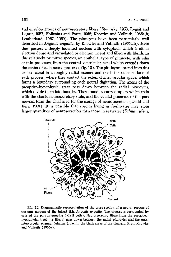
the center of each neural process ( Fig. 10). The pituicytes extend from this
central canal in a roughly radial manner and reach the outer surface of
each process, where they contact the external intervascular space, which
forms a boundary surrounding each neural digitation. The axons of the
preoptico-hypophysial tract pass down between the radial pituicytes,
which divide them into bundles. These bundles carry droplets which stain
with the classic neurosecretory stain, and the caudal processes of the pars
nervosa form the chief area for the storage of neurosecretion (Dodd and
Kerr, 1963). It is possible that species living in freshwater may store
larger quantities of neurosecretion than those in seawater (Salmo irideus,
- MSH
Pituicyte
ns
Fil
Channel
W
Fig. 10. Diagrammatic representation of the cross section of a neural process of
the pars nervosa of the teleost fish, Anguillu anguillu. The process is surrounded by
cells of the pars intermedia (MSH cells). Neurosecretory fibers from the preoptimhypophysial tract (ns fibers) pass down between the radial pituicytes and the outer
intervascular channel (channel), i.e., in the black areas of the diagram. From Knowles
and Vollrath ( 1965a).
A. M. PERKS
and envelop groups of neurosecretory fibers (Stutinsky, 1953; Legait and
Legait, 1957; Follenius and Porte, 1962; Knowles and Vollrath, 1965a,b;
Leatherland, 1967, 1Q69). The pituicytes have been particularly well
described in Anguilla anguilla, by Knowles and Vollrath ( 1965a,b). Here
they possess a deeply indented nucleus with cytoplasm which is either
electron dense and vacuolated or electron lucent and filled with fibrilli. In
this relatively primitive species, an epithelial type of pituicyte, with cilia
or thin processes, lines the central ventricular canal which extends down
the center of each neural process ( Fig. 10). The pituicytes extend from this
central canal in a roughly radial manner and reach the outer surface of
each process, where they contact the external intervascular space, which
forms a boundary surrounding each neural digitation. The axons of the
preoptico-hypophysial tract pass down between the radial pituicytes,
which divide them into bundles. These bundles carry droplets which stain
with the classic neurosecretory stain, and the caudal processes of the pars
nervosa form the chief area for the storage of neurosecretion (Dodd and
Kerr, 1963). It is possible that species living in freshwater may store
larger quantities of neurosecretion than those in seawater (Salmo irideus,
- MSH
Pituicyte
ns
Fil
Channel
W
Fig. 10. Diagrammatic representation of the cross section of a neural process of
the pars nervosa of the teleost fish, Anguillu anguillu. The process is surrounded by
cells of the pars intermedia (MSH cells). Neurosecretory fibers from the preoptimhypophysial tract (ns fibers) pass down between the radial pituicytes and the outer
intervascular channel (channel), i.e., in the black areas of the diagram. From Knowles
and Vollrath ( 1965a).