

2. THE NEUROHYPOPHYSIS
157
I
1 0
2
12
3
II
4
13
5
7
6
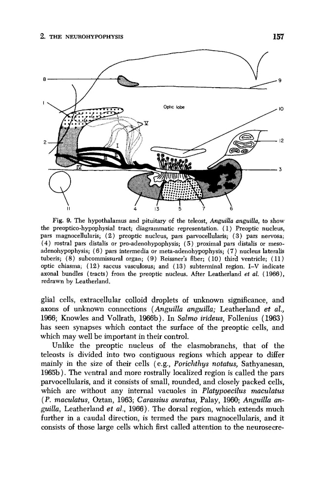
Fig. 9. The hypothalamus and pituitary of the teleost, Anguilla anguilla, to show
the preoptico-hypophysial tract; diagrammatic representation. ( 1 ) Preoptic nucleus,
pars magnocellularis; ( 2 ) preoptic nucleus, pars parvocellularis; ( 3 ) pars nervosa;
( 4 ) rostra1 pars distalis or pro-adenohypophysis; ( 5 ) ,proximal pars distalis or mesoadenohypophysis; ( 6 ) pars intermedia or meta-adenohypophysis; ( 7 ) nucleus lateralis
tuberis; ( 8 ) subcommissural organ; ( 9 ) Reissner’s fiber; ( 10) third ventricle; ( 11 )
optic chiasma; (12) saccus vasculosus; and (13) subterminal region. I-V indicate
axonal bundles (tracts) from the preoptic nucleus. After Leatherland et al. ( 19&3),
redrawn by Leatherland.
glial cells, extracellular colloid droplets of unknown significance, and
axons of unknown connections ( Anguilla anguilla; Leatherland et al.,
1966; Knowles and Vollrath, 1966b). In Salmo irideus, Follenius (1963)
has seen synapses which contact the surface of the preoptic cells, and
which may well be important in their control.
Unlike the preoptic nucleus of the elasmobranchs, that of the
teleosts is divided into two contiguous regions which appear to differ
mainly in the size of their cells (e.g., Porichthys notatus, Sathyanesan,
196513 ) . The ventral and more rostrally localized region is called the pars
parvocellularis, and it consists of small, rounded, and closely packed cells,
which are without any internal vacuoles in Platypoecilus maculatus
( P . maculutus, Oztan, 1963; Carassius auratus, Palay, 1960; AnguiZZu anguilla, Leatherland et al., 1966). The dorsal region, which extends much
further in a caudal direction, is termed the pars magnocellularis, and it
consists of those large cells which first called attention to the neurosecre-
157
I
1 0
2
12
3
II
4
13
5
7
6
Fig. 9. The hypothalamus and pituitary of the teleost, Anguilla anguilla, to show
the preoptico-hypophysial tract; diagrammatic representation. ( 1 ) Preoptic nucleus,
pars magnocellularis; ( 2 ) preoptic nucleus, pars parvocellularis; ( 3 ) pars nervosa;
( 4 ) rostra1 pars distalis or pro-adenohypophysis; ( 5 ) ,proximal pars distalis or mesoadenohypophysis; ( 6 ) pars intermedia or meta-adenohypophysis; ( 7 ) nucleus lateralis
tuberis; ( 8 ) subcommissural organ; ( 9 ) Reissner’s fiber; ( 10) third ventricle; ( 11 )
optic chiasma; (12) saccus vasculosus; and (13) subterminal region. I-V indicate
axonal bundles (tracts) from the preoptic nucleus. After Leatherland et al. ( 19&3),
redrawn by Leatherland.
glial cells, extracellular colloid droplets of unknown significance, and
axons of unknown connections ( Anguilla anguilla; Leatherland et al.,
1966; Knowles and Vollrath, 1966b). In Salmo irideus, Follenius (1963)
has seen synapses which contact the surface of the preoptic cells, and
which may well be important in their control.
Unlike the preoptic nucleus of the elasmobranchs, that of the
teleosts is divided into two contiguous regions which appear to differ
mainly in the size of their cells (e.g., Porichthys notatus, Sathyanesan,
196513 ) . The ventral and more rostrally localized region is called the pars
parvocellularis, and it consists of small, rounded, and closely packed cells,
which are without any internal vacuoles in Platypoecilus maculatus
( P . maculutus, Oztan, 1963; Carassius auratus, Palay, 1960; AnguiZZu anguilla, Leatherland et al., 1966). The dorsal region, which extends much
further in a caudal direction, is termed the pars magnocellularis, and it
consists of those large cells which first called attention to the neurosecre-