

2. THE NEUROHYPOPHYSIS
149
VI. BONY FISH: THE CHONDROSTEI
A. The Structure of the Neurohypophysis of the Chondrostei
The Chondrostei, which include the sturgeons and the paddlefish,
represent a distinct branch of the evolutionary tree (Dodd and Ken,
1963). However, they may be closer to the main trunk of vertebrate
evolution than the teleost fish (Polenov, 1966). Their pituitaries are more
primitive than those of the teleosts, and they are more closely allied to
those of Polypterm and the brachiopterygians (Jasinski, 1964). In recent
years, their neurohypophysial system has been described by Polenov and
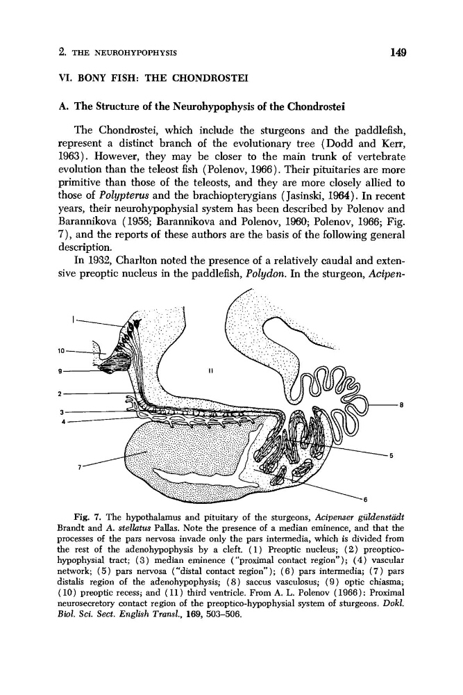
Barannikova (1958; Barannikova and Polenov, 1960; Polenov, 1966; Fig.
7 ) , and the reports of these authors are the basis of the following general
description.
In 1932, Charlton noted the presence of a relatively caudal and extensive preoptic nucleus in the paddlefish, Polydon. In the sturgeon, Acipen- 8
. 5
Fig. 7. The hypothalamus and pituitary of the sturgeons, Acipenser guldenstadt
Brandt and A. stelhtus Pallas. Note the presence of a median eminence, and that the
processes of the pars nervosa invade only the pars intermedia, which is divided from
the rest of the adenohypophysis by a cleft. ( 1 ) Preoptic nucleus; (2) preopticohypophysial tract; ( 3 ) median eminence ( “proximal contact region”); ( 4 ) vascular
network; ( 5 ) pars nervosa (“distal contact region”); ( 6 ) pars intermedia; ( 7 ) pars
distalis region of the adenohypophysis; ( 8 ) saccus vasculosus; ( 9 ) optic chiasma;
(10) preoptic recess; and (11) third ventricle. From A. L. Polenov (1966): Proximal
neurosecretory contact region of the preoptico-hypophysial system of sturgeons. Dokl.
Biol. Sci. Sect. English Transl., 169, 503-506.
149
VI. BONY FISH: THE CHONDROSTEI
A. The Structure of the Neurohypophysis of the Chondrostei
The Chondrostei, which include the sturgeons and the paddlefish,
represent a distinct branch of the evolutionary tree (Dodd and Ken,
1963). However, they may be closer to the main trunk of vertebrate
evolution than the teleost fish (Polenov, 1966). Their pituitaries are more
primitive than those of the teleosts, and they are more closely allied to
those of Polypterm and the brachiopterygians (Jasinski, 1964). In recent
years, their neurohypophysial system has been described by Polenov and
Barannikova (1958; Barannikova and Polenov, 1960; Polenov, 1966; Fig.
7 ) , and the reports of these authors are the basis of the following general
description.
In 1932, Charlton noted the presence of a relatively caudal and extensive preoptic nucleus in the paddlefish, Polydon. In the sturgeon, Acipen- 8
. 5
Fig. 7. The hypothalamus and pituitary of the sturgeons, Acipenser guldenstadt
Brandt and A. stelhtus Pallas. Note the presence of a median eminence, and that the
processes of the pars nervosa invade only the pars intermedia, which is divided from
the rest of the adenohypophysis by a cleft. ( 1 ) Preoptic nucleus; (2) preopticohypophysial tract; ( 3 ) median eminence ( “proximal contact region”); ( 4 ) vascular
network; ( 5 ) pars nervosa (“distal contact region”); ( 6 ) pars intermedia; ( 7 ) pars
distalis region of the adenohypophysis; ( 8 ) saccus vasculosus; ( 9 ) optic chiasma;
(10) preoptic recess; and (11) third ventricle. From A. L. Polenov (1966): Proximal
neurosecretory contact region of the preoptico-hypophysial system of sturgeons. Dokl.
Biol. Sci. Sect. English Transl., 169, 503-506.