

2. THE NEUROHYPOPHYSIS
143
f
1
n
v
:::i:i:i:. :. :::::::i:
.:... . .:.:.:.:.. . .
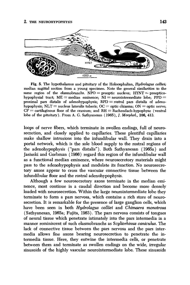
Fig. 5. The hypothalamus and pituitary of the Holocephalian, Hydrolagus colliei;
median sagittal section from a young specimen. Note the general similarities to the
same region of the elasmobranchs. NPO = preoptic nucleus; HYNT = preopticohypophysial tract; ME = median eminence; NI = neurointermediate lobe; PPD =
proximal pars distalis of adenohypophysis; RPD = rostral pars distalis of adenohypophysis; NLT = nucleus lateralis tuberis; OC = optic chiasma; ON = optic nerve;
CF = cartilaginous floor of the cranium; and RH = Rachendach-hypophyse (ventral
lobe of the pituitary). From A. G. Sathyanesan (1965), J. Morphol., 106, 413.
loops of nerve fibers, which terminate in swollen endings, full of neurosecretion, and closely applied to capillaries. These plentiful capillaries
make shallow intrusions into the infundibular wall. They drain into a
portal network, which is the sole blood supply to the rostral regions of
the adenohypophysis (“pars distalis”), Both Sathyanesan ( 196!5a) and
Jashiski and Gorbman (1966) regard this region of the infundibular wall
as a functional median eminence, where neurosecretory materials might
pass to the adenohypophysis and modulate its function. No neurosecretory axons appear to cross the vascular connective tissue between the
infundibular floor and the rostral adenohypophysis.
Although a few neurosecretory axons terminate in the median eminence, most continue in a caudal direction and become more densely
loaded with neurosecretion. Within the large neurointermediate lobe they
terminate to form a pars nervosa, which contains a rich store of neurosecretion. It is remarkable for the presence of large ganglion cells, which
have been seen in both Hydrolugus colliei and Chimaera monstrosa
( Sathyanesan, 1965a; Fujita, 1963). The pars nervosa consists of tongues
of neural tissue which penetrate intimately into the pars intermedia in a
manner reminiscent of such elasmobranchs as Scyliorhinus caniculus. The
lack of connective tissue between the pars nervosa and the pars intermedia allows fine axons bearing neurosecretion to penetrate the intermedia tissue. Here, they entwine the intermedia cells, or penetrate
between them and terminate as swollen endings on the wide, irregular
sinusoids of the highly vascular neurointermediate lobe. These sinusoids
143
f
1
n
v
:::i:i:i:. :. :::::::i:
.:... . .:.:.:.:.. . .
Fig. 5. The hypothalamus and pituitary of the Holocephalian, Hydrolagus colliei;
median sagittal section from a young specimen. Note the general similarities to the
same region of the elasmobranchs. NPO = preoptic nucleus; HYNT = preopticohypophysial tract; ME = median eminence; NI = neurointermediate lobe; PPD =
proximal pars distalis of adenohypophysis; RPD = rostral pars distalis of adenohypophysis; NLT = nucleus lateralis tuberis; OC = optic chiasma; ON = optic nerve;
CF = cartilaginous floor of the cranium; and RH = Rachendach-hypophyse (ventral
lobe of the pituitary). From A. G. Sathyanesan (1965), J. Morphol., 106, 413.
loops of nerve fibers, which terminate in swollen endings, full of neurosecretion, and closely applied to capillaries. These plentiful capillaries
make shallow intrusions into the infundibular wall. They drain into a
portal network, which is the sole blood supply to the rostral regions of
the adenohypophysis (“pars distalis”), Both Sathyanesan ( 196!5a) and
Jashiski and Gorbman (1966) regard this region of the infundibular wall
as a functional median eminence, where neurosecretory materials might
pass to the adenohypophysis and modulate its function. No neurosecretory axons appear to cross the vascular connective tissue between the
infundibular floor and the rostral adenohypophysis.
Although a few neurosecretory axons terminate in the median eminence, most continue in a caudal direction and become more densely
loaded with neurosecretion. Within the large neurointermediate lobe they
terminate to form a pars nervosa, which contains a rich store of neurosecretion. It is remarkable for the presence of large ganglion cells, which
have been seen in both Hydrolugus colliei and Chimaera monstrosa
( Sathyanesan, 1965a; Fujita, 1963). The pars nervosa consists of tongues
of neural tissue which penetrate intimately into the pars intermedia in a
manner reminiscent of such elasmobranchs as Scyliorhinus caniculus. The
lack of connective tissue between the pars nervosa and the pars intermedia allows fine axons bearing neurosecretion to penetrate the intermedia tissue. Here, they entwine the intermedia cells, or penetrate
between them and terminate as swollen endings on the wide, irregular
sinusoids of the highly vascular neurointermediate lobe. These sinusoids