

2. THE NEUROHYPOPHYSIS
129
6
A. Hypothetic
B. Squalus
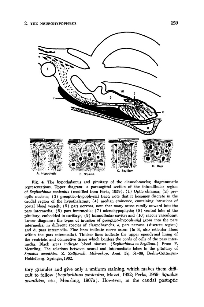
Fig. 4. The hypothalamus and pituitary of the elasmobranchs; diagrammatic
representations. Upper diagram: a parasagittal section of the infundibular region
of Scyliorhinus caniculus (modified from Perks, 1959). ( 1 ) Optic chiasma; ( 2 ) preoptic nucleus; (3) preoptico-hypophysial tract; note that it becomes discrete in the
caudal region of the hypothalamus; ( 4 ) median eminence, containing intrusions of
portal blood vessels; ( 5 ) pars nervosa, note that many axons ramify onward into the
pars intermedia; (6) pars intermedia; ( 7 ) adenohypophysis; (8) ventral lobe of the
pituitary, embedded in cartilage; ( 9 ) infundibular cavity; and ( 10) saccus vasculosus.
Lower diagrams: the types of invasion of preoptico-hypophysial axons into the pars
intermedia, in different species of elasmobranchs. a, pars nervosa (discrete region)
and b, pars intermedia. Fine lines indicate nerve axons (in B, also reticular fibers
within the pars intermedia). Thicker lines indicate the upper ependymal lining of
the ventricle, and connective tissue which borders the cords of cells of the pars intermedia. Black areas indicate blood sinuses. ( Scyliorhinus = Scyllium. ) From P.
Meurling, The relations between neural and intermediate lobes in the pituitary of
Squalus acanthias. Z. Zellforsch. Mikroskop. Anat. 58, 51-69, Berlin-GijttingenHeidelberg: Springer, 1962.
tory granules and give only a uniform staining, which makes them d a -
cult to follow (Scyliorhinus caniculus, Mazzi, 1952; Perks, 1959; Squalus
acanthias, etc., Meurling, 1967a). However, in the caudal postoptic
129
6
A. Hypothetic
B. Squalus
Fig. 4. The hypothalamus and pituitary of the elasmobranchs; diagrammatic
representations. Upper diagram: a parasagittal section of the infundibular region
of Scyliorhinus caniculus (modified from Perks, 1959). ( 1 ) Optic chiasma; ( 2 ) preoptic nucleus; (3) preoptico-hypophysial tract; note that it becomes discrete in the
caudal region of the hypothalamus; ( 4 ) median eminence, containing intrusions of
portal blood vessels; ( 5 ) pars nervosa, note that many axons ramify onward into the
pars intermedia; (6) pars intermedia; ( 7 ) adenohypophysis; (8) ventral lobe of the
pituitary, embedded in cartilage; ( 9 ) infundibular cavity; and ( 10) saccus vasculosus.
Lower diagrams: the types of invasion of preoptico-hypophysial axons into the pars
intermedia, in different species of elasmobranchs. a, pars nervosa (discrete region)
and b, pars intermedia. Fine lines indicate nerve axons (in B, also reticular fibers
within the pars intermedia). Thicker lines indicate the upper ependymal lining of
the ventricle, and connective tissue which borders the cords of cells of the pars intermedia. Black areas indicate blood sinuses. ( Scyliorhinus = Scyllium. ) From P.
Meurling, The relations between neural and intermediate lobes in the pituitary of
Squalus acanthias. Z. Zellforsch. Mikroskop. Anat. 58, 51-69, Berlin-GijttingenHeidelberg: Springer, 1962.
tory granules and give only a uniform staining, which makes them d a -
cult to follow (Scyliorhinus caniculus, Mazzi, 1952; Perks, 1959; Squalus
acanthias, etc., Meurling, 1967a). However, in the caudal postoptic