

2. THE NEUROHYPOPHYSIS
117
19
20
1 0 1 1 12 13 14
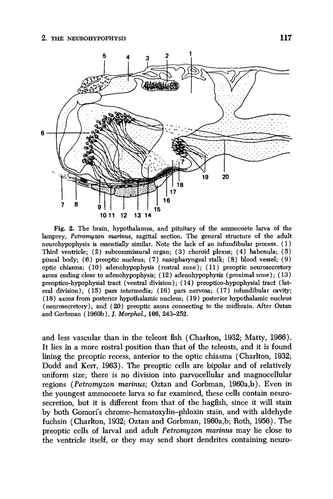
Fig. 2. The brain, hypothalamus, and pituitary of the ammocoete larva of the
lamprey, Petromyzon marinus, sagittal section. The general structure of the adult
neurohypophysis is essentially similar. Note the lack of an infundibular process. ( 1 )
Third ventricle; ( 2 ) subcommissural organ; ( 3 ) choroid plexus; ( 4 ) habenula; ( 5 )
pineal body; ( 6 ) preoptic nucleus; ( 7 ) nasopharyngeal stalk; ( 8) blood vessel; ( 9 )
optic chiasma; ( 10) adenohypophysis (rostral zone); ( 11) preoptic neurosecretory
axons ending close to adenohypophysis; ( 12) adenohypophysis (proximal zone); (13)
preoptico-hypophysial tract (ventral division) ; ( 14) preoptico-hypophysial tract (lateral division); ( 15) pars intermedia; (16) pars nervosa; ( 17) infundibular cavity;
(18) axons from posterior hypothalamic nucleus; ( 1 9 ) posterior hypothalamic nucleus
(neurosecretory); and ( 2 0 ) preoptic axons connecting to the midbrain. After Oztan
and Gorbman (1960b), J. Morphol., 106,243-252.
and less vascular than in the teleost fish (Charlton, 1932; Matty, 1966).
It lies in a more rostral position than that of the teleosts, and it is found
lining the preoptic recess, anterior to the optic chiasma (Charlton, 1932;
Dodd and Kerr, 1963). The preoptic cells are bipolar and of relatively
uniform size; there is no division into parvocellular and magnocellular
regions ( Petromyzon marinus; Oztan and Gorbman, 1960a,b). Even in
the youngest ammocoete larva so far examined, these cells contain neurosecretion, but it is different from that of the hagfish, since it will stain
by both Gomori's chrome-hematoxylin-phloxin stain, and with aldehyde
fuchsin (Charlton, 1932; Oztan and Gorbman, 1960a,b; Roth, 1956). The
preoptic cells of larval and adult Petromyzon marinus may lie close to
the ventricle itself, or they may send short dendrites containing neuro-
117
19
20
1 0 1 1 12 13 14
Fig. 2. The brain, hypothalamus, and pituitary of the ammocoete larva of the
lamprey, Petromyzon marinus, sagittal section. The general structure of the adult
neurohypophysis is essentially similar. Note the lack of an infundibular process. ( 1 )
Third ventricle; ( 2 ) subcommissural organ; ( 3 ) choroid plexus; ( 4 ) habenula; ( 5 )
pineal body; ( 6 ) preoptic nucleus; ( 7 ) nasopharyngeal stalk; ( 8) blood vessel; ( 9 )
optic chiasma; ( 10) adenohypophysis (rostral zone); ( 11) preoptic neurosecretory
axons ending close to adenohypophysis; ( 12) adenohypophysis (proximal zone); (13)
preoptico-hypophysial tract (ventral division) ; ( 14) preoptico-hypophysial tract (lateral division); ( 15) pars intermedia; (16) pars nervosa; ( 17) infundibular cavity;
(18) axons from posterior hypothalamic nucleus; ( 1 9 ) posterior hypothalamic nucleus
(neurosecretory); and ( 2 0 ) preoptic axons connecting to the midbrain. After Oztan
and Gorbman (1960b), J. Morphol., 106,243-252.
and less vascular than in the teleost fish (Charlton, 1932; Matty, 1966).
It lies in a more rostral position than that of the teleosts, and it is found
lining the preoptic recess, anterior to the optic chiasma (Charlton, 1932;
Dodd and Kerr, 1963). The preoptic cells are bipolar and of relatively
uniform size; there is no division into parvocellular and magnocellular
regions ( Petromyzon marinus; Oztan and Gorbman, 1960a,b). Even in
the youngest ammocoete larva so far examined, these cells contain neurosecretion, but it is different from that of the hagfish, since it will stain
by both Gomori's chrome-hematoxylin-phloxin stain, and with aldehyde
fuchsin (Charlton, 1932; Oztan and Gorbman, 1960a,b; Roth, 1956). The
preoptic cells of larval and adult Petromyzon marinus may lie close to
the ventricle itself, or they may send short dendrites containing neuro-