

2. THE NEUROHYPOPHYSIS
115
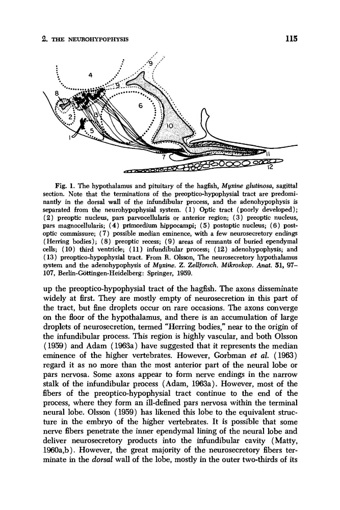
Fig. 1. The hypothalamus and pituitary of the hagfish, Myxine glutinosu, sagittal
section. Note that the terminations of the preoptico-hypophysial tract are predominantly in the dorsal wall of the infundibular process, and the adenohypophysis is
separated from the neurohypophysial system. ( 1 ) Optic tract ( poorly developed ) ;
( 2 ) preoptic nucleus, pars parvocellularis or anterior region; (3) preoptic nucleus,
pars magnocellularis; (4) primordium hippocampi; (5) postoptic nucleus; (6) postoptic commissure; (7) possible median eminence, with a few neurosecretory endings
(Herring bodies); ( 8 ) preoptic recess; ( 9 ) areas of remnants of buried ependymal
cells; (10) third ventricle; (11) infundibular process; (12) adenohypophysis; and
( 13) preoptico-hypophysial tract. From R. Olsson, The neurosecretory hypothalamus
system and the adenohypophysis of Myxine. Z. Zellforsch. Mikroskop. Anat. 51, 97107, Berlin-Gattingen-Heidelberg: Springer, 1959.
up the preoptico-hypophysial tract of the hagfish. The axons disseminate
widely at first. They are mostly empty of neurosecretion in this part of
the tract, but fine droplets occur on rare occasions. The axons converge
on the floor of the hypothalamus, and there is an accumulation of large
droplets of neurosecretion, termed “Herring bodies,” near to the origin of
the infundibular process. This region is highly vascular, and both Olsson
(1959) and Adam (1963a) have suggested that it represents the median
eminence of the higher vertebrates. However, Gorbman et a2. (1963)
regard it as no more than the most anterior part of the neural lobe or
pars nervosa. Some axons appear to form nerve endings in the narrow
stalk of the infundibular process (Adam, 1963a). However, most of the
fibers of the preoptico-hypophysial tract continue to the end of the
process, where they form an ill-defined pars nervosa within the terminal
neural lobe. Olsson (1959) has likened this lobe to the equivalent structure in the embryo of the higher vertebrates. It is possible that some
nerve fibers penetrate the inner ependymal lining of the neural lobe and
deliver neurosecretory products into the infundibular cavity ( Matty,
1960a,b). However, the great majority of the neurosecretory fibers terminate in the dorsal wall of the lobe, mostly in the outer two-thirds of its
115
Fig. 1. The hypothalamus and pituitary of the hagfish, Myxine glutinosu, sagittal
section. Note that the terminations of the preoptico-hypophysial tract are predominantly in the dorsal wall of the infundibular process, and the adenohypophysis is
separated from the neurohypophysial system. ( 1 ) Optic tract ( poorly developed ) ;
( 2 ) preoptic nucleus, pars parvocellularis or anterior region; (3) preoptic nucleus,
pars magnocellularis; (4) primordium hippocampi; (5) postoptic nucleus; (6) postoptic commissure; (7) possible median eminence, with a few neurosecretory endings
(Herring bodies); ( 8 ) preoptic recess; ( 9 ) areas of remnants of buried ependymal
cells; (10) third ventricle; (11) infundibular process; (12) adenohypophysis; and
( 13) preoptico-hypophysial tract. From R. Olsson, The neurosecretory hypothalamus
system and the adenohypophysis of Myxine. Z. Zellforsch. Mikroskop. Anat. 51, 97107, Berlin-Gattingen-Heidelberg: Springer, 1959.
up the preoptico-hypophysial tract of the hagfish. The axons disseminate
widely at first. They are mostly empty of neurosecretion in this part of
the tract, but fine droplets occur on rare occasions. The axons converge
on the floor of the hypothalamus, and there is an accumulation of large
droplets of neurosecretion, termed “Herring bodies,” near to the origin of
the infundibular process. This region is highly vascular, and both Olsson
(1959) and Adam (1963a) have suggested that it represents the median
eminence of the higher vertebrates. However, Gorbman et a2. (1963)
regard it as no more than the most anterior part of the neural lobe or
pars nervosa. Some axons appear to form nerve endings in the narrow
stalk of the infundibular process (Adam, 1963a). However, most of the
fibers of the preoptico-hypophysial tract continue to the end of the
process, where they form an ill-defined pars nervosa within the terminal
neural lobe. Olsson (1959) has likened this lobe to the equivalent structure in the embryo of the higher vertebrates. It is possible that some
nerve fibers penetrate the inner ependymal lining of the neural lobe and
deliver neurosecretory products into the infundibular cavity ( Matty,
1960a,b). However, the great majority of the neurosecretory fibers terminate in the dorsal wall of the lobe, mostly in the outer two-thirds of its