

1. ANATOMY AND PHYSIOLOGY OF THE CENTRAL NERVOUS SYSTEM
45
D
D
A
P
A
P
V
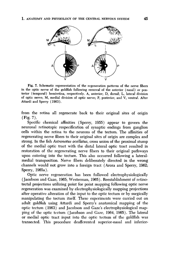
Fig. 7. Schematic representation of the regeneration patterns of the nerve fibers
in the optic nerve of the goldfish following removal of the anterior (nasal) or posterior ( temporal) hemiretina, respectively. A, anterior; D, dorsal; L, lateral division
of optic nerve; M, medial division of optic nerve; P, posterior; and V, ventral. After
Attardi and Sperry ( 1963).
from the retina all regenerate back to their original sites of origin
(Fig. 7 ) .
Specific chemical affinities (Sperry, 1955) appear to govern the
neuronal retinotopic respecification of synaptic endings from ganglion
cells within the retina to the neurons of the tectum. The affinities of
regenerating nerve fibers to their original sites of origin are complex and
strong. In the fish Astronotus ocellatus, cross union of the proximal stump
of the medial optic tract with the distal lateral optic tract resulted in
restoration of the regenerating nerve fibers to their original pathways
upon entering into the tectum. This also occurred following a Jateralmedial transposition. Nerve fibers deliberately directed in the wrong
channels would not grow into a foreign tract (Arora and Sperry, 1962;
Sperry, 1965a).
Optic nerve regeneration has been followed electrophysiologically
(Jacobson and Gaze, 1965; Westerman, 1965). Reestablishment of retinotectal projections utilizing point for point mapping following optic nerve
regeneration was examined by electrophysiologically mapping projections
after operative alteration of the input to the optic tectum or by surgically
manipulating the tectum itself, These experiments were carried out on
adult goldfish using Attardi and Sperry’s anatomical mapping of the
optic tectum ( 1963) and Jacobson and Gaze’s electrophysiological mapping of the optic tcctum (Jacobson and Gaze, 1961, 1965). The lateral
or medial optic tract input into the optic tectum of the goldfish was
transected. This procedure deafferentcd superior-nasal and inferior-
45
D
D
A
P
A
P
V
Fig. 7. Schematic representation of the regeneration patterns of the nerve fibers
in the optic nerve of the goldfish following removal of the anterior (nasal) or posterior ( temporal) hemiretina, respectively. A, anterior; D, dorsal; L, lateral division
of optic nerve; M, medial division of optic nerve; P, posterior; and V, ventral. After
Attardi and Sperry ( 1963).
from the retina all regenerate back to their original sites of origin
(Fig. 7 ) .
Specific chemical affinities (Sperry, 1955) appear to govern the
neuronal retinotopic respecification of synaptic endings from ganglion
cells within the retina to the neurons of the tectum. The affinities of
regenerating nerve fibers to their original sites of origin are complex and
strong. In the fish Astronotus ocellatus, cross union of the proximal stump
of the medial optic tract with the distal lateral optic tract resulted in
restoration of the regenerating nerve fibers to their original pathways
upon entering into the tectum. This also occurred following a Jateralmedial transposition. Nerve fibers deliberately directed in the wrong
channels would not grow into a foreign tract (Arora and Sperry, 1962;
Sperry, 1965a).
Optic nerve regeneration has been followed electrophysiologically
(Jacobson and Gaze, 1965; Westerman, 1965). Reestablishment of retinotectal projections utilizing point for point mapping following optic nerve
regeneration was examined by electrophysiologically mapping projections
after operative alteration of the input to the optic tectum or by surgically
manipulating the tectum itself, These experiments were carried out on
adult goldfish using Attardi and Sperry’s anatomical mapping of the
optic tectum ( 1963) and Jacobson and Gaze’s electrophysiological mapping of the optic tcctum (Jacobson and Gaze, 1961, 1965). The lateral
or medial optic tract input into the optic tectum of the goldfish was
transected. This procedure deafferentcd superior-nasal and inferior-