

9. AIR BREATHING IN FISHES
407
surface and gulps air which is placed as a bubble between the tongue and
the buccal roof. When stimulated to breath air by low O2 tension in
the water, the buccal epithelial capillaries promptly dilate and become
engorged with blood, giving the epithelium a corrugated lunglike appearance, bright red from a proliferous circulation. This change in perfusion
occurs within a few minutes and is totally absent in related fishes which
are purely aquatic breathers (Todd and Ebeling, 19SS). The close
coupling between mechanisms controlling breathing and vasomotor responses seems basic to very diverse types of respiratory organs ranging
from the gills in fishes to the lungs of both lower and higher vertebrates.
Only in the lungfishes among air-breathing fishes has the vascular
circuit to the air-breathing organ gained enough anatomical separation
from the systemic circuit to allow separate selective perfusion of the
two circuits.
During rest in aerated water, the lung in Neoceratodus is of no
I
50
100
1 50
200
210
pMo2
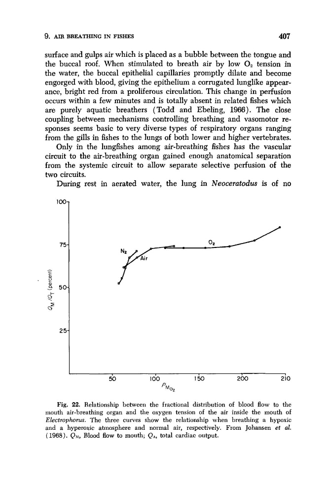
Fig. 22. Relationship between the fractional distribution of blood flow to the
mouth air-breathing organ and the oxygen tension of the air inside the mouth of
Electrophorus. The three curves show the relationship when breathing a hypoxic
and a hyperoxic atmosphere and normal air, respectively. From Johansen et al.
( 1968). QJI, Blood flow to mouth; QA, total cardiac output.
407
surface and gulps air which is placed as a bubble between the tongue and
the buccal roof. When stimulated to breath air by low O2 tension in
the water, the buccal epithelial capillaries promptly dilate and become
engorged with blood, giving the epithelium a corrugated lunglike appearance, bright red from a proliferous circulation. This change in perfusion
occurs within a few minutes and is totally absent in related fishes which
are purely aquatic breathers (Todd and Ebeling, 19SS). The close
coupling between mechanisms controlling breathing and vasomotor responses seems basic to very diverse types of respiratory organs ranging
from the gills in fishes to the lungs of both lower and higher vertebrates.
Only in the lungfishes among air-breathing fishes has the vascular
circuit to the air-breathing organ gained enough anatomical separation
from the systemic circuit to allow separate selective perfusion of the
two circuits.
During rest in aerated water, the lung in Neoceratodus is of no
I
50
100
1 50
200
210
pMo2
Fig. 22. Relationship between the fractional distribution of blood flow to the
mouth air-breathing organ and the oxygen tension of the air inside the mouth of
Electrophorus. The three curves show the relationship when breathing a hypoxic
and a hyperoxic atmosphere and normal air, respectively. From Johansen et al.
( 1968). QJI, Blood flow to mouth; QA, total cardiac output.