

8. THE REGULATION OF BREATHING
307
Phy’
/ / / 1
Si hy
Hy hy
Ce
Add
md2
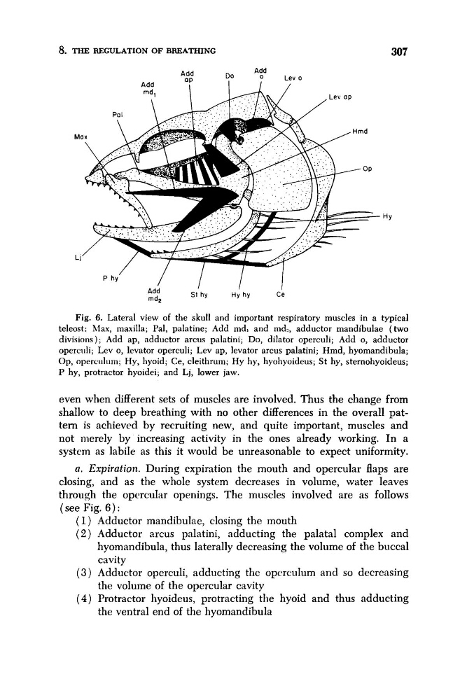
Fig. 6. Lateral view of the skull and important respiratory muscles in a typical
teleost: hlax, maxilla; Pal, palatine; Add md, and md,, adductor mandibulae (two
divisions); Add ap, adductor arcus palatini; Do, dilator operculi; Add 0, adductor
operculi; Lev 0, lcvator operculi; Lev ap, levator arcus palatini; Hmd, hyomandibula;
Op, operculum; Hy, hyoid; Ce, cleithrum; Hy hy, hyohyoideus; St hy, sternohyoideus;
P hy, protractor hyoidei; and Lj, lower jaw.
even when different sets of muscles are involved. Thus the change from
shallow to deep breathing with no other differences in the overall pattern is achieved by recruiting new, and quite important, muscles and
not merely by increasing activity in the ones already working. In a
system as labile as this it would be unreasonable to expect uniformity.
a. Expiration. During expiration the mouth and opercular flaps are
closing, and as the whole system decreases in volume, water leaves
through the opcrcular openings. The muscles involved are as follows
(see Fig. 6 ) :
(1) Adductor mandibulae, closing the mouth
( 2 ) Adductor arcus pnlatini, adducting the palatal complex and
hyomandibula, thus laterally decreasing the volume of the buccal
cavity
( 3 ) Adductor operculi, adducting the opcwdum and so decreasing
the volume of the opercular cavity
(4) Protractor hyoideus, protracting the hyoid and thus adducting
the ventral end of the hyomandibula
307
Phy’
/ / / 1
Si hy
Hy hy
Ce
Add
md2
Fig. 6. Lateral view of the skull and important respiratory muscles in a typical
teleost: hlax, maxilla; Pal, palatine; Add md, and md,, adductor mandibulae (two
divisions); Add ap, adductor arcus palatini; Do, dilator operculi; Add 0, adductor
operculi; Lev 0, lcvator operculi; Lev ap, levator arcus palatini; Hmd, hyomandibula;
Op, operculum; Hy, hyoid; Ce, cleithrum; Hy hy, hyohyoideus; St hy, sternohyoideus;
P hy, protractor hyoidei; and Lj, lower jaw.
even when different sets of muscles are involved. Thus the change from
shallow to deep breathing with no other differences in the overall pattern is achieved by recruiting new, and quite important, muscles and
not merely by increasing activity in the ones already working. In a
system as labile as this it would be unreasonable to expect uniformity.
a. Expiration. During expiration the mouth and opercular flaps are
closing, and as the whole system decreases in volume, water leaves
through the opcrcular openings. The muscles involved are as follows
(see Fig. 6 ) :
(1) Adductor mandibulae, closing the mouth
( 2 ) Adductor arcus pnlatini, adducting the palatal complex and
hyomandibula, thus laterally decreasing the volume of the buccal
cavity
( 3 ) Adductor operculi, adducting the opcwdum and so decreasing
the volume of the opercular cavity
(4) Protractor hyoideus, protracting the hyoid and thus adducting
the ventral end of the hyomandibula