

1. ANATOMY AND PHYSIOLOGY OF THE CENTRAL NERVOUS SYSTEM
5
the dendrites and the occasional lamination with free ribosomes into
Nissl bodies. Many dendrites contain regularly, longitudinally oriented
neurotubules or canaliculi of indefinite length and about 200 A in diameter as well as neurofilaments. In the goldfish, dendritic neurotubules
can be observed in regular laminated patterns (Fig. 1). Dendrites also
possess many spinous projections (dendritic spines) which are sites of
increased neuronal area for synaptic contact ( axodendritic synapses).
Neurofilaments are a conspicuous element of axoplasm. Smooth endoplasmic reticulum is common and rough endoplasmic reticulum is absent.
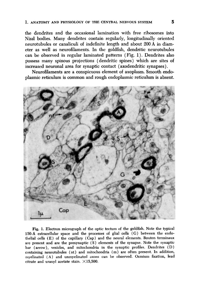
Fig. 1. Electron micrograph of the optic tectum of the goldfish. Note the typical
150-A extracellular space and the processes of glial cells ( G ) between the endothelial cells ( E ) of the capillary ( C a p ) and the neural elements. Bouton terminaux
are present and are the presynaptic ( S ) elements of the synapse. Note the synaptic
bar (arrow), vesicles, and mitochondria in the synaptic profiles. Dendrites ( D )
containing neurotubules ( n t ) and mitochondria ( m ) are often present. In addition,
myelinated ( A ) and unmyelinated axons can be observed. Osmium fixation, lead
citrate and uranyl acetate stain. X 15,500.
5
the dendrites and the occasional lamination with free ribosomes into
Nissl bodies. Many dendrites contain regularly, longitudinally oriented
neurotubules or canaliculi of indefinite length and about 200 A in diameter as well as neurofilaments. In the goldfish, dendritic neurotubules
can be observed in regular laminated patterns (Fig. 1). Dendrites also
possess many spinous projections (dendritic spines) which are sites of
increased neuronal area for synaptic contact ( axodendritic synapses).
Neurofilaments are a conspicuous element of axoplasm. Smooth endoplasmic reticulum is common and rough endoplasmic reticulum is absent.
Fig. 1. Electron micrograph of the optic tectum of the goldfish. Note the typical
150-A extracellular space and the processes of glial cells ( G ) between the endothelial cells ( E ) of the capillary ( C a p ) and the neural elements. Bouton terminaux
are present and are the presynaptic ( S ) elements of the synapse. Note the synaptic
bar (arrow), vesicles, and mitochondria in the synaptic profiles. Dendrites ( D )
containing neurotubules ( n t ) and mitochondria ( m ) are often present. In addition,
myelinated ( A ) and unmyelinated axons can be observed. Osmium fixation, lead
citrate and uranyl acetate stain. X 15,500.