

G W M E CAMPBELL
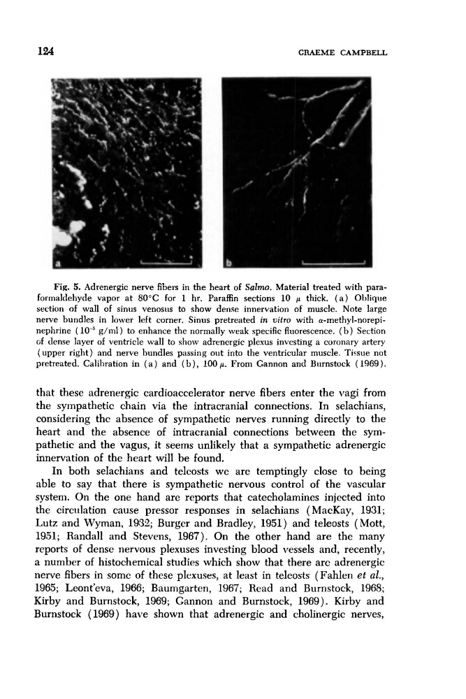
Fig. 5. Adrenergic nerve fibers in the heart of Salmo. Material treated with paraformaldehyde vapor at 80°C for 1 hr. Paraffin sections 10 p thick. ( a ) Oblique
section of wall of sinus venosiis to show dense innervation of muscle. Note large
nerve bundles in lower left corner. Sinus pretreated in uitm with a-methyl-norepinephrine (
g/nil) to enhance the normally weak specific fluorescence. ( b ) Section
c;f dense layer of ventricle wall to show adrenergic plexus investing a coronary artery
(upper right) and nerve bundles passing out into the ventricular muscle. Ticsue not
pretreated. Calibration in ( a ) and ( b ) , 100 p. From Cannon and Burnstock ( 1969).
that these adrenergic cardioaccelerator nerve fibers enter the vagi from
the sympathetic chain via the intracranial connections. In selachians,
considering the absence of sympathetic nerves running directly to the
heart and the absence of intracranial connections between the sympathetic and the vagus, it seems unlikely that a sympathetic adrenergic
innervation of the heart will be found.
In both selachians and telcosts we are temptingly close to being
able to say that there is sympathetic nervous control of the vascular
system. On the one hand are reports that catecholamines injected into
the circulation cause pressor responses in selachians ( MacKay, 1931;
Lutz and Wyman, 1932; Burger and Bradley, 1951) and teleosts (Mott,
1951; Randall and Stevens, 1967). On the other hand are the many
reports of dense nervous plexuses investing blood vessels and, recently,
a number of histochemical studies which show that there arc adrenergic
nerve fibers in some of these plexuses, at least in teleosts (Fahlen et al.,
1965; Leont’eva, 1966; Baumgarten, 1967; Read and Burnstock, 1968;
Kirby and Burnstock, 1969; Gannon and Burnstock, 1969). Kirby and
Burnstock (1969) have shown that adrenergic and cholinergic nerves,
Fig. 5. Adrenergic nerve fibers in the heart of Salmo. Material treated with paraformaldehyde vapor at 80°C for 1 hr. Paraffin sections 10 p thick. ( a ) Oblique
section of wall of sinus venosiis to show dense innervation of muscle. Note large
nerve bundles in lower left corner. Sinus pretreated in uitm with a-methyl-norepinephrine (
g/nil) to enhance the normally weak specific fluorescence. ( b ) Section
c;f dense layer of ventricle wall to show adrenergic plexus investing a coronary artery
(upper right) and nerve bundles passing out into the ventricular muscle. Ticsue not
pretreated. Calibration in ( a ) and ( b ) , 100 p. From Cannon and Burnstock ( 1969).
that these adrenergic cardioaccelerator nerve fibers enter the vagi from
the sympathetic chain via the intracranial connections. In selachians,
considering the absence of sympathetic nerves running directly to the
heart and the absence of intracranial connections between the sympathetic and the vagus, it seems unlikely that a sympathetic adrenergic
innervation of the heart will be found.
In both selachians and telcosts we are temptingly close to being
able to say that there is sympathetic nervous control of the vascular
system. On the one hand are reports that catecholamines injected into
the circulation cause pressor responses in selachians ( MacKay, 1931;
Lutz and Wyman, 1932; Burger and Bradley, 1951) and teleosts (Mott,
1951; Randall and Stevens, 1967). On the other hand are the many
reports of dense nervous plexuses investing blood vessels and, recently,
a number of histochemical studies which show that there arc adrenergic
nerve fibers in some of these plexuses, at least in teleosts (Fahlen et al.,
1965; Leont’eva, 1966; Baumgarten, 1967; Read and Burnstock, 1968;
Kirby and Burnstock, 1969; Gannon and Burnstock, 1969). Kirby and
Burnstock (1969) have shown that adrenergic and cholinergic nerves,