

3. AUTONOMIC NERVOUS SYSTEMS
113
There is, however, general agreement that there are neither organized
sympathetic chains nor strictly segmental sympathetic ganglia in cyclostomes. There are collections of ganglion cells aIong the cardinal veins
in the abdominal cavity of lampreys which may represent sympathetic
ganglia. In Lampetru, visceral branches of the spinal nerves run directly
to the region of blood vessels, the kidneys, and the gonads. In posterior
regions there is a spinal outflow to the cloaca, rectum, and ureters, with
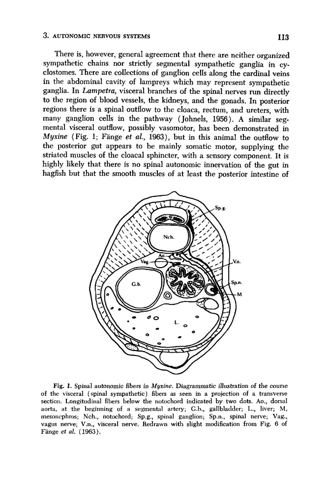
many ganglion cells in the pathway (Johnels, 1956). A similar segmental visceral outflow, possibly vasomotor, has been demonstrated in
Myxine (Fig. 1; Fange et al., 1963), but in this animal the outflow to
the posterior gut appears to be mainly somatic motor, supplying the
striated muscles of the cloaca1 sphincter, with a sensory component. It is
highly likely that there is no spinal autonomic innervation of the gut in
hagfish but that the smooth muscles of at least the posterior intestine of
Fig. 1. Spinal autonomic fibers in Myxine. Diagrammatic illustration of the course
of the visceral (spinal sympathetic) fibers as seen in a projection of a transverse
section. Longitudinal fibers below the notochord indicated by two dots. Ao., dorsal
aorta, at the beginning of a segmental artery; G.h., gallbladder; L., liver; M,
niesoncphros; Nch., notochord; Sp.g., spinal ganglion; Sp.n., spinal nerve; Vag.,
vagus nerve; V.n., visceral nerve. Redrawn with slight modification from Fig. 6 of
Fange et al. (1963).
113
There is, however, general agreement that there are neither organized
sympathetic chains nor strictly segmental sympathetic ganglia in cyclostomes. There are collections of ganglion cells aIong the cardinal veins
in the abdominal cavity of lampreys which may represent sympathetic
ganglia. In Lampetru, visceral branches of the spinal nerves run directly
to the region of blood vessels, the kidneys, and the gonads. In posterior
regions there is a spinal outflow to the cloaca, rectum, and ureters, with
many ganglion cells in the pathway (Johnels, 1956). A similar segmental visceral outflow, possibly vasomotor, has been demonstrated in
Myxine (Fig. 1; Fange et al., 1963), but in this animal the outflow to
the posterior gut appears to be mainly somatic motor, supplying the
striated muscles of the cloaca1 sphincter, with a sensory component. It is
highly likely that there is no spinal autonomic innervation of the gut in
hagfish but that the smooth muscles of at least the posterior intestine of
Fig. 1. Spinal autonomic fibers in Myxine. Diagrammatic illustration of the course
of the visceral (spinal sympathetic) fibers as seen in a projection of a transverse
section. Longitudinal fibers below the notochord indicated by two dots. Ao., dorsal
aorta, at the beginning of a segmental artery; G.h., gallbladder; L., liver; M,
niesoncphros; Nch., notochord; Sp.g., spinal ganglion; Sp.n., spinal nerve; Vag.,
vagus nerve; V.n., visceral nerve. Redrawn with slight modification from Fig. 6 of
Fange et al. (1963).