

378
N. BALAKRISHNAN NAIR AND M. SARASW-ATHY
CO
EC
K A U V R PA
C
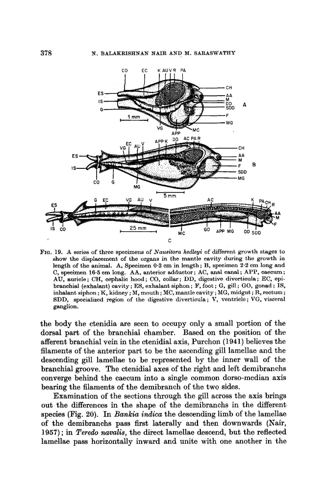
FIG. 19. A series of three specimens of Nauitora hedleyi of different growth stages to
show the displacement of the organs in the mantle cavity during the growth in
length of the animal. A, Specimen 0.3 cm in length; B, specimen 2.2 cm long and
C, specimen 16.5 cm long. AA, anterior adductor ; AC, anal canal ; APP, caecum :
AU, auricle ; CH, cephalic hood ; CO, collar ; DD, digestive diverticula ; EC, epibranchial (exhalant) cavity ; ES, exhalant siphon ; F, foot ; G, gill ; GO, gonad : IS,
inhalant siphon ; K, kidney ; M, mouth ; MC, mantle cavity ; MG, midgut ; R, rectum ;
SDD, speoialized region of the digestive diverticula ; V, ventricle ; VG, visceral
ganglion.
the body the ctenidia are seen to occupy only a small portion of the
dorsal part of the branchial chamber. Based on the position of the
afferent branchial vein in the ctenidial axis, Purchon (1941) believes the
filaments of the anterior part to be the ascending gill lamellae and the
descending gill lamellae to be represented by the inner wall of the
branchial groove. The ctenidial axes of the right and left demibranchs
converge behind the caecum into a single common dorso-median axis
bearing the filaments of the demibranch of the two sides.
Examination of the sections through the gill across the axis brings
out the differences in the shape of the demibranchs in the different
species (Fig. 20). In Bankia indica the descending limb of the lamellae
of the demibranchs pass first laterally and then downwards (Nair,
1957) ; in Teredo nawalis, the direct lamellae descend, but the reflected
lamellae pass horizontally inward and unite with one another in the
N. BALAKRISHNAN NAIR AND M. SARASW-ATHY
CO
EC
K A U V R PA
C
FIG. 19. A series of three specimens of Nauitora hedleyi of different growth stages to
show the displacement of the organs in the mantle cavity during the growth in
length of the animal. A, Specimen 0.3 cm in length; B, specimen 2.2 cm long and
C, specimen 16.5 cm long. AA, anterior adductor ; AC, anal canal ; APP, caecum :
AU, auricle ; CH, cephalic hood ; CO, collar ; DD, digestive diverticula ; EC, epibranchial (exhalant) cavity ; ES, exhalant siphon ; F, foot ; G, gill ; GO, gonad : IS,
inhalant siphon ; K, kidney ; M, mouth ; MC, mantle cavity ; MG, midgut ; R, rectum ;
SDD, speoialized region of the digestive diverticula ; V, ventricle ; VG, visceral
ganglion.
the body the ctenidia are seen to occupy only a small portion of the
dorsal part of the branchial chamber. Based on the position of the
afferent branchial vein in the ctenidial axis, Purchon (1941) believes the
filaments of the anterior part to be the ascending gill lamellae and the
descending gill lamellae to be represented by the inner wall of the
branchial groove. The ctenidial axes of the right and left demibranchs
converge behind the caecum into a single common dorso-median axis
bearing the filaments of the demibranch of the two sides.
Examination of the sections through the gill across the axis brings
out the differences in the shape of the demibranchs in the different
species (Fig. 20). In Bankia indica the descending limb of the lamellae
of the demibranchs pass first laterally and then downwards (Nair,
1957) ; in Teredo nawalis, the direct lamellae descend, but the reflected
lamellae pass horizontally inward and unite with one another in the