

264
THE BIOLOGY O F EUPHAUSIIDS
An excretory organ is present in the base of each of the antennae
and is supplied with blood via branches of the two arteriae laterales
(Fig. 97). This organ, like those of other crustaceans, can be divided
histologically into three regions, the end sac, the urinary duct, and the
urethra. The end sac, oval in shape and connected to the urinary duct
by an opening, has a network of blood lacunae inside it. The opening
between the sac and the urinary duct is guarded by a ring of six cells
forming a valve to prevent excretory products flowing back into the
sac; no sphincter muscle is present. The urinary duct, forming a ring
around the end sac, is not of uniform diameter but has a wide part
which forms a reservoir for the excretory products. According to Raab,
the urinary duct not only carries the excretory products to the outside
but its walls also have an excretory function. The external opening of
the gland is at a small papilla on the ventral side of the base of the
antenna. Raab describes the different types of epithelia in the various
regions of the organ while Zimmer (1913) and Chun (1896) describe the
excretory organs of Euphausia superba and Stylocheiron sp. respectively
but not fully as neither author noticed an end sac. Nothing is known
about the functioning of this organ in euphausiids because investigations of the water and salt balance of these animals have not been made
as yet. The only study of excretion in euphausiids is that of Conover
and Corner (1968) who found that Meganyctiphanes norvegica excreted
0.19-0.60 pg nitrogenlmg dry wt/day a t 3"-5"C.
Descriptions of the gross morphology of the nervous system of
euphausiids are available (Sars, 1885; Zimmer, 1913; Raab, 1915) but
very little is known about the detailed structure of the cerebral ganglion,
the ventral nerve cord, or the stomatogastric nervous system. The
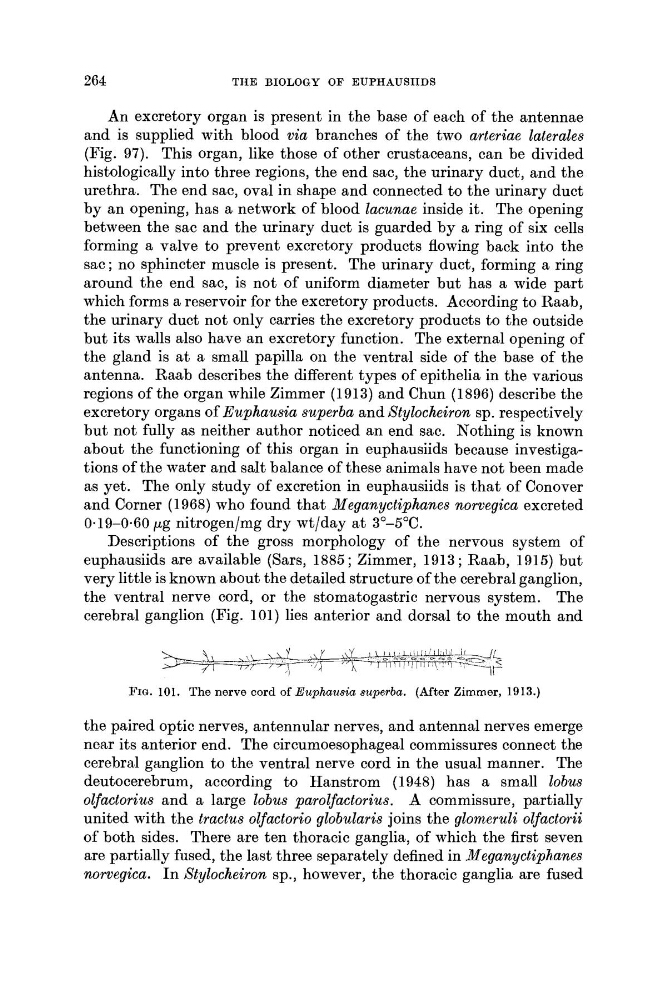
cerebral ganglion (Fig. 101) lies anterior and dorsal to the mouth and
FIG. 101. The nerve cord of Euphausia superba. (After Zimmer, 1913.)
the paired optic nerves, antennular nerves, and antenna1 nerves emerge
near its anterior end. The circumoesophageal commissures connect the
cerebral ganglion to the ventral nerve cord in the usual manner. The
deutocerebrum, according to Hanstrom (1948) has a small lobus
oljactorius and a large lobus paroljactorius. A commissure, partially
united with the tractus oljactorio gbobularis joins the glomeruli ouactorii
of both sides. There are ten thoracic ganglia, of which the first seven
are partially fused, the last three separately defined in Meganyctiphanes
norvegica. I n Stylocheiron sp., however, the thoracic ganglia are fused
THE BIOLOGY O F EUPHAUSIIDS
An excretory organ is present in the base of each of the antennae
and is supplied with blood via branches of the two arteriae laterales
(Fig. 97). This organ, like those of other crustaceans, can be divided
histologically into three regions, the end sac, the urinary duct, and the
urethra. The end sac, oval in shape and connected to the urinary duct
by an opening, has a network of blood lacunae inside it. The opening
between the sac and the urinary duct is guarded by a ring of six cells
forming a valve to prevent excretory products flowing back into the
sac; no sphincter muscle is present. The urinary duct, forming a ring
around the end sac, is not of uniform diameter but has a wide part
which forms a reservoir for the excretory products. According to Raab,
the urinary duct not only carries the excretory products to the outside
but its walls also have an excretory function. The external opening of
the gland is at a small papilla on the ventral side of the base of the
antenna. Raab describes the different types of epithelia in the various
regions of the organ while Zimmer (1913) and Chun (1896) describe the
excretory organs of Euphausia superba and Stylocheiron sp. respectively
but not fully as neither author noticed an end sac. Nothing is known
about the functioning of this organ in euphausiids because investigations of the water and salt balance of these animals have not been made
as yet. The only study of excretion in euphausiids is that of Conover
and Corner (1968) who found that Meganyctiphanes norvegica excreted
0.19-0.60 pg nitrogenlmg dry wt/day a t 3"-5"C.
Descriptions of the gross morphology of the nervous system of
euphausiids are available (Sars, 1885; Zimmer, 1913; Raab, 1915) but
very little is known about the detailed structure of the cerebral ganglion,
the ventral nerve cord, or the stomatogastric nervous system. The
cerebral ganglion (Fig. 101) lies anterior and dorsal to the mouth and
FIG. 101. The nerve cord of Euphausia superba. (After Zimmer, 1913.)
the paired optic nerves, antennular nerves, and antenna1 nerves emerge
near its anterior end. The circumoesophageal commissures connect the
cerebral ganglion to the ventral nerve cord in the usual manner. The
deutocerebrum, according to Hanstrom (1948) has a small lobus
oljactorius and a large lobus paroljactorius. A commissure, partially
united with the tractus oljactorio gbobularis joins the glomeruli ouactorii
of both sides. There are ten thoracic ganglia, of which the first seven
are partially fused, the last three separately defined in Meganyctiphanes
norvegica. I n Stylocheiron sp., however, the thoracic ganglia are fused