

234
THE BIOLOGY OF EUPHAUSIIDS
These two ganglia have a very rich supply of blood and vessels pass
from one to the other. The optic artery, now in the dorsal region of the
eye, supplies by branches the lamina ganglionaris and the region
between it and the basement membrane. This region below the basement membrane is very well supplied with blood vessels which tend to
run parallel to each other round the membrane from the dorsal to the
ventral side of the eye. Consequently, sections of the eye can be cut in
which the majority of vessels are obvious as transverse sections or in
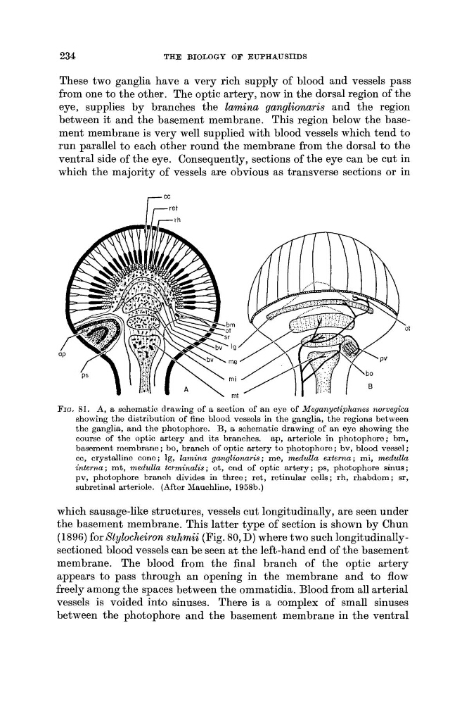
FIG. 81. A, a schematic drawing of a section of an eye of Meganyctiphanes norvegica
showing the distribution of fine blood vessels in the ganglia, the regions between
the ganglia, and the photophore. B, a schematic drawing of an eye showing the
course of the optic artery and its branches. ap, arteriole in photophore; bm,
basement membrane ; bo, branch of optic artery to photophore ; bv, blood vessel ;
cc, crystalline cone ; lg, lamina ganglionaris ; me, medulla externa ; mi, medulla
interna; mt, medulla terminalis; ot, end of optic artery; ps, photophore sinus;
pv, photophore branch divides in three; ret, retinular cells; rh, rhabdom; sr,
subretinal arteriole. (After Mauchline, 1958b.)
which sausage-like structures, vessels cut longitudinally, are seen under
the basement membrane. This latter type of section is shown by Chun
(1896) for Xtylocheiron suhmii (Fig. 80, D) where two such longitudinallysectioned blood vessels can be seen a t the left-hand end of the basement
membrane. The blood from the final branch of the optic artery
appears t o pass through an opening in the membrane and to flow
freely among the spaces between the ommatidia. Blood from all arterial
vessels is voided into sinuses. There is a complex of small sinuses
between the photophore and the basement membrane in the ventral
THE BIOLOGY OF EUPHAUSIIDS
These two ganglia have a very rich supply of blood and vessels pass
from one to the other. The optic artery, now in the dorsal region of the
eye, supplies by branches the lamina ganglionaris and the region
between it and the basement membrane. This region below the basement membrane is very well supplied with blood vessels which tend to
run parallel to each other round the membrane from the dorsal to the
ventral side of the eye. Consequently, sections of the eye can be cut in
which the majority of vessels are obvious as transverse sections or in
FIG. 81. A, a schematic drawing of a section of an eye of Meganyctiphanes norvegica
showing the distribution of fine blood vessels in the ganglia, the regions between
the ganglia, and the photophore. B, a schematic drawing of an eye showing the
course of the optic artery and its branches. ap, arteriole in photophore; bm,
basement membrane ; bo, branch of optic artery to photophore ; bv, blood vessel ;
cc, crystalline cone ; lg, lamina ganglionaris ; me, medulla externa ; mi, medulla
interna; mt, medulla terminalis; ot, end of optic artery; ps, photophore sinus;
pv, photophore branch divides in three; ret, retinular cells; rh, rhabdom; sr,
subretinal arteriole. (After Mauchline, 1958b.)
which sausage-like structures, vessels cut longitudinally, are seen under
the basement membrane. This latter type of section is shown by Chun
(1896) for Xtylocheiron suhmii (Fig. 80, D) where two such longitudinallysectioned blood vessels can be seen a t the left-hand end of the basement
membrane. The blood from the final branch of the optic artery
appears t o pass through an opening in the membrane and to flow
freely among the spaces between the ommatidia. Blood from all arterial
vessels is voided into sinuses. There is a complex of small sinuses
between the photophore and the basement membrane in the ventral