

306
ELVEZIO GHIRARDELLI
volume they are almost never a t the same level. As a consequence, the
ovaries develop unequally, the one whose ampullae is more cephalic
being the longer.
The intestine, in the portion between the two ovaries, has a sinuous
course because the ampullae, owing to their width, protrude beyond
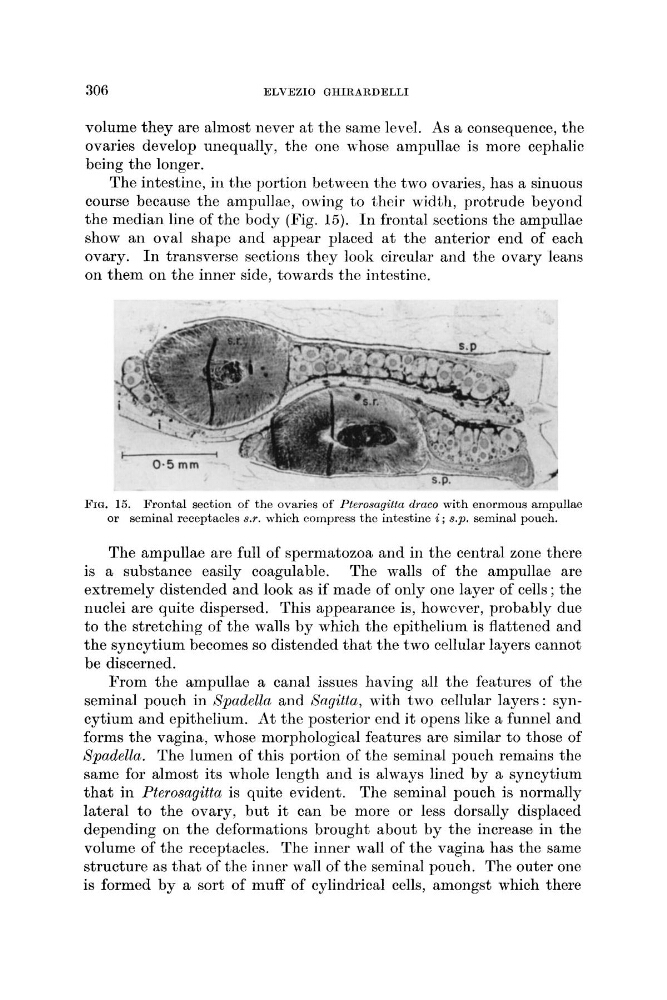
the median line of the body (Fig. 15). I n frontal sections the ampullae
show an oval shape and appear placed a t the anterior end of each
ovary. I n transverse sections they look circular and the ovary leans
on them on the inner side, towards the intestine.
FIG. 15. Frontal section of the ovaries of Pterosagittn clrnco with enormous ampullae
or seminal receptacles s.r. which cornpress the intestine i ; s.p. seminal pouch.
The ampullae are full of spermatozoa and in the central zone there
is a substance easily coagulable. The wdls of the ampullae are
extremely distended and look as if made of only one layer of cells ; the
nuclei are quite dispersed. This appearance is, however, probably due
to the stretching of the walls by which the epithelium is flattened and
the syncytium becomes so distended that the two cellular layers cannot
be discerned.
From the ampullae a canal issues having all the features of the
seminal pouch in Spadella and Bagitta, with two cellular layers: syncytium and epithelium. At the posterior end i t opens like a funnel and
forms the vagina, whose morphological features are similar to those of
Spadella. The lumen of this portion of the seminal pouch remains the
same for almost its whole length and is always lined by a syncytium
that in Pterosagitta is quite evident. The seminal pouch is normally
lateral to the ovary, but it can be more or less dorsally displaced
depending on the deformations brought about by the increase in the
volume of the receptacles. The inner wall of the vagina has the same
structure as that of the inner wall of the seminal pouch. The outer one
is formed by a sort of muff of cylindrical cells, amongst which there
ELVEZIO GHIRARDELLI
volume they are almost never a t the same level. As a consequence, the
ovaries develop unequally, the one whose ampullae is more cephalic
being the longer.
The intestine, in the portion between the two ovaries, has a sinuous
course because the ampullae, owing to their width, protrude beyond
the median line of the body (Fig. 15). I n frontal sections the ampullae
show an oval shape and appear placed a t the anterior end of each
ovary. I n transverse sections they look circular and the ovary leans
on them on the inner side, towards the intestine.
FIG. 15. Frontal section of the ovaries of Pterosagittn clrnco with enormous ampullae
or seminal receptacles s.r. which cornpress the intestine i ; s.p. seminal pouch.
The ampullae are full of spermatozoa and in the central zone there
is a substance easily coagulable. The wdls of the ampullae are
extremely distended and look as if made of only one layer of cells ; the
nuclei are quite dispersed. This appearance is, however, probably due
to the stretching of the walls by which the epithelium is flattened and
the syncytium becomes so distended that the two cellular layers cannot
be discerned.
From the ampullae a canal issues having all the features of the
seminal pouch in Spadella and Bagitta, with two cellular layers: syncytium and epithelium. At the posterior end i t opens like a funnel and
forms the vagina, whose morphological features are similar to those of
Spadella. The lumen of this portion of the seminal pouch remains the
same for almost its whole length and is always lined by a syncytium
that in Pterosagitta is quite evident. The seminal pouch is normally
lateral to the ovary, but it can be more or less dorsally displaced
depending on the deformations brought about by the increase in the
volume of the receptacles. The inner wall of the vagina has the same
structure as that of the inner wall of the seminal pouch. The outer one
is formed by a sort of muff of cylindrical cells, amongst which there