

SOiIIE ASPECTS O F THE BIOLOGY O F THE CHAETOGNATHS
287
concavity facing the inner ring. At least two layers of cells usually
form the outer ring; a similar disposition may not be present in the
inner ring, which might be formed by one layer only.
The inner ring is consequently less developed, and this is quite the
contrary of what one may see in Spadella. This disposition is, however,
not a constant one: it may vary even in the same corona, where at
some points the margins may be bent and not at others. There is no
clear separation, in Pterosagitta draco, between the inner and the outer
ring. One can only say that the nuclei of the cells of the middle line,
which is interposed between the two concentric rings, lie somewhat
deeper than the nuclei of the lateral portions. All the cells of the corona
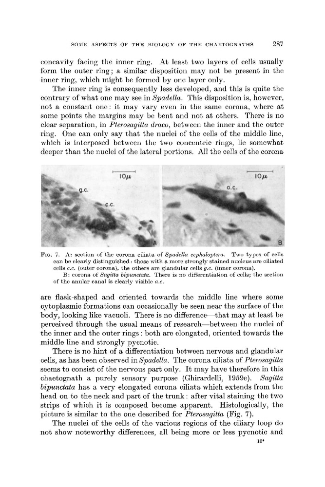
FIG. 7. A: section of the corona ciliata of Spadelln cephaloptern. Two types of cells
can be clearly distinguished : those with a more strongly stained nucleus are ciliated
cells C.C. (outer corona). the others are glandular cells g.c. (inner corona).
B: corona of Sagitta bipunctata. There is no differentiation of cells; the section
of the anular canal is clearly visible a.c.
are flask-shaped and oriented towards the middle line where some
cytoplasmic formations can occasionally be seen near the surface of the
body, looking like vacuoli. There is no difference-that may at least be
perceived through the usual means of research-between the nuclei of
the inner and the outer rings : both are elongated, oriented towards the
middle line and strongly pycnotic.
There is no hint of a differentiation between nervous and glandular
cells, as has been observed in Spudella. The corona ciliata of Pterosagitta
seems to consist of the nervous part only. It may have therefore in this
chaetognath a purely sensory purpose (Ghirardelli, 1959e). Sagitta
bipunctata has a very elongated corona ciliata which extends from the
head on to the neck and part of the trunk : after vital staining the two
strips of which it is composed become apparent. Histologically, the
picture is similar to the one described for Pterosagitta (Fig. 7).
The nuclei of the cells of the various regions of the ciliary loop do
not show noteworthy differences, all being more or less pycnotic and
10'
287
concavity facing the inner ring. At least two layers of cells usually
form the outer ring; a similar disposition may not be present in the
inner ring, which might be formed by one layer only.
The inner ring is consequently less developed, and this is quite the
contrary of what one may see in Spadella. This disposition is, however,
not a constant one: it may vary even in the same corona, where at
some points the margins may be bent and not at others. There is no
clear separation, in Pterosagitta draco, between the inner and the outer
ring. One can only say that the nuclei of the cells of the middle line,
which is interposed between the two concentric rings, lie somewhat
deeper than the nuclei of the lateral portions. All the cells of the corona
FIG. 7. A: section of the corona ciliata of Spadelln cephaloptern. Two types of cells
can be clearly distinguished : those with a more strongly stained nucleus are ciliated
cells C.C. (outer corona). the others are glandular cells g.c. (inner corona).
B: corona of Sagitta bipunctata. There is no differentiation of cells; the section
of the anular canal is clearly visible a.c.
are flask-shaped and oriented towards the middle line where some
cytoplasmic formations can occasionally be seen near the surface of the
body, looking like vacuoli. There is no difference-that may at least be
perceived through the usual means of research-between the nuclei of
the inner and the outer rings : both are elongated, oriented towards the
middle line and strongly pycnotic.
There is no hint of a differentiation between nervous and glandular
cells, as has been observed in Spudella. The corona ciliata of Pterosagitta
seems to consist of the nervous part only. It may have therefore in this
chaetognath a purely sensory purpose (Ghirardelli, 1959e). Sagitta
bipunctata has a very elongated corona ciliata which extends from the
head on to the neck and part of the trunk : after vital staining the two
strips of which it is composed become apparent. Histologically, the
picture is similar to the one described for Pterosagitta (Fig. 7).
The nuclei of the cells of the various regions of the ciliary loop do
not show noteworthy differences, all being more or less pycnotic and
10'