

by the snail’s proboscis during feeding. According to Cole, 0. eulimoides
commonly occurs in pairs, but three may be found within the same
pocket.
Description. Body 3-6 mm long ; shell white, opaque, (with faint
orange or purplish tinge a t apical end of living specimens) elongate
conical, covered with minute yellow specks and pale yellow line
longitudinally oriented along each side ; with fine and close-set microscopical spiral striae; apex tapering to a blunt point; six t o seven
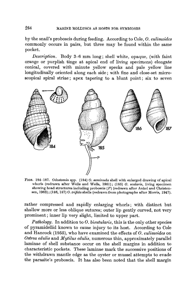
FIGS. 184-187. Odostomia spp. (184) 0. aelninuda shell with enlarged drawing of apical
whorls (redrawn after Wells and Wells, 1961); (186) 0. scalaris, living specimen
showing head structures including proboscis ( P ) (redrawn after Ankel and Christensen, 1963) ; (186,187) 0. trifida shells (redrawn from photographs after Morris, 1947).
rather compressed and rapidly enlarging whorls ; with distinct but
shallow more or less oblique sutures; outer lip gently curved, not very
prominent ; inner lip very slight, limited to upper part.
Pathology. I n addition to 0. bisutularis, this is the only other species
of pyramidellid known t o cause injury t o its host. According to Cole
and Hancock (1955), who have examined the effects of 0. eulimoides on
Ostrea edulis and Mytilus edulis, numerous thin, approximately parallel
laminae of shell substance occur on the shell margins in addition t o
characteristic pockets. These laminae mark the successive positions of
the withdrawn mantle edge as the oyster or mussel attempts to evade
the parasite’s proboscis. It has also been noted that the shell margin
commonly occurs in pairs, but three may be found within the same
pocket.
Description. Body 3-6 mm long ; shell white, opaque, (with faint
orange or purplish tinge a t apical end of living specimens) elongate
conical, covered with minute yellow specks and pale yellow line
longitudinally oriented along each side ; with fine and close-set microscopical spiral striae; apex tapering to a blunt point; six t o seven
FIGS. 184-187. Odostomia spp. (184) 0. aelninuda shell with enlarged drawing of apical
whorls (redrawn after Wells and Wells, 1961); (186) 0. scalaris, living specimen
showing head structures including proboscis ( P ) (redrawn after Ankel and Christensen, 1963) ; (186,187) 0. trifida shells (redrawn from photographs after Morris, 1947).
rather compressed and rapidly enlarging whorls ; with distinct but
shallow more or less oblique sutures; outer lip gently curved, not very
prominent ; inner lip very slight, limited to upper part.
Pathology. I n addition to 0. bisutularis, this is the only other species
of pyramidellid known t o cause injury t o its host. According to Cole
and Hancock (1955), who have examined the effects of 0. eulimoides on
Ostrea edulis and Mytilus edulis, numerous thin, approximately parallel
laminae of shell substance occur on the shell margins in addition t o
characteristic pockets. These laminae mark the successive positions of
the withdrawn mantle edge as the oyster or mussel attempts to evade
the parasite’s proboscis. It has also been noted that the shell margin