

5. THE PHYLUM PROTOZOA
163
spore measures 2.42-4-20 p (average 3.09 p) x 2-14-3.26 p (average
2.58 p), without projections ; sporocysts 7.014 p (average 9 p ) in diameter, each containing twenty to fifty spores.
Life cycle. Although Wood and Andrews (1962) have given certain
aspects of the life cycle of M . costalis, its complete life cycle remains
undetermined.
According to Wood and Andrews, the earliest stage observed in
the oyster is a small multinucleated plasmodium which is more or
less irregular in outline. Its cell membrane is at first definite but becomes
less definite with age. As each plasmodium increases in size, its nuclei
multiply in number and the cytoplasm becomes vacuolated. The multinucleated plasmodium eventually cleaves into uninucleated portions
each of which develops into a characteristic spore. At this point the
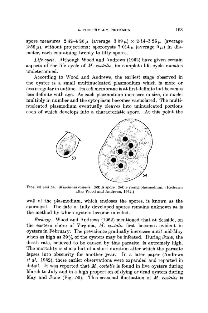
FIQS. 53 and 54. Minchinia costalis. (53) A spore; (54) a young plasmodium. (Redrawn
after Wood and Andrews, 1962.)
wall of the plasmodium, which encloses the spores, is known as the
sporocyst. The fate of fully developed spores remains unknown as is
the method by which oysters become infected.
Ecology. Wood and Andrews (1962) mentioned that a t Seaside, on
the eastern shore of Virginia, M . costalis first becomes evident in
oysters in February. The prevalence gradually increases until mid-May
when as high as 39% of the oysters may be infected. During June, the
death rate, believed to be caused by this parasite, is extremely high.
The mortality is sharp but of a short duration after which the parasite
lapses into obscurity for another year. In a later paper (Andrews
et al., 1962), these earlier observations were expanded and reported in
detail. It was reported that M . costalis is found in live oysters during
March to July and in a high proportion of dying or dead oysters during
May and June (Fig. 55). This seasonal fluctuation of M . costalis is
163
spore measures 2.42-4-20 p (average 3.09 p) x 2-14-3.26 p (average
2.58 p), without projections ; sporocysts 7.014 p (average 9 p ) in diameter, each containing twenty to fifty spores.
Life cycle. Although Wood and Andrews (1962) have given certain
aspects of the life cycle of M . costalis, its complete life cycle remains
undetermined.
According to Wood and Andrews, the earliest stage observed in
the oyster is a small multinucleated plasmodium which is more or
less irregular in outline. Its cell membrane is at first definite but becomes
less definite with age. As each plasmodium increases in size, its nuclei
multiply in number and the cytoplasm becomes vacuolated. The multinucleated plasmodium eventually cleaves into uninucleated portions
each of which develops into a characteristic spore. At this point the
FIQS. 53 and 54. Minchinia costalis. (53) A spore; (54) a young plasmodium. (Redrawn
after Wood and Andrews, 1962.)
wall of the plasmodium, which encloses the spores, is known as the
sporocyst. The fate of fully developed spores remains unknown as is
the method by which oysters become infected.
Ecology. Wood and Andrews (1962) mentioned that a t Seaside, on
the eastern shore of Virginia, M . costalis first becomes evident in
oysters in February. The prevalence gradually increases until mid-May
when as high as 39% of the oysters may be infected. During June, the
death rate, believed to be caused by this parasite, is extremely high.
The mortality is sharp but of a short duration after which the parasite
lapses into obscurity for another year. In a later paper (Andrews
et al., 1962), these earlier observations were expanded and reported in
detail. It was reported that M . costalis is found in live oysters during
March to July and in a high proportion of dying or dead oysters during
May and June (Fig. 55). This seasonal fluctuation of M . costalis is