

126
MARINE MOLLUSCS AS HOSTS FOR SYMBIOSES
Another manifestation of trematode infection in molluscs was
originally reported by Willey a n d Gross (1957). During the process of
seeking some visible characteristic which would permit the separation
of uninfected Littorina littorea from those infected with Cryptocotyle
lingua, these investigators noticed that the foot of parasitized periwinkles consistently portray a dark yellow brown color while the foot
of uninfected ones remains white. I n addition to C. lingua, it has been
found that periwinkles infected with two other species of trematodes
(unidentified) also portray the characteristic brown color. I n attempts
to identify the pigment(s), the following characteristics have been
400
4 5 0
500
550
600
6 5 0
700
WAVELENGTH^^)
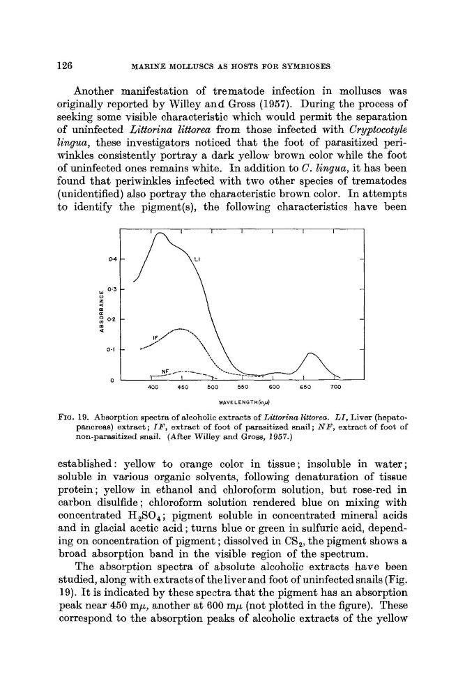
PIQ. 19. Absorption spectra of alcoholic extracts of Littorina Zittorecc. LI, Liver (hepatopancreas) extract ; I F , extract of foot of parasitized snail ; N P , extract of foot of
non-parasitized snail. (After Willey and Gross, 1957.)
established: yellow to orange color in tissue; insoluble in water;
soluble in various organic solvents, following denaturation of tissue
protein ; yellow in ethanol and chloroform solution, but rose-red in
carbon disulfide ; chloroform solution rendered blue on mixing with
concentrated H,SO, ; pigment soluble in concentrated mineral acids
and in glacial acetic acid ; turns blue or green in sulfuric acid, depending on concentration of pigment ; dissolved in CS,, the pigment shows a
broad absorption band in the visible region of the spectrum.
The absorption spectra of absolute alcoholic extracts have been
studied, along with extracts of the liver and foot of uninfected snails (Fig.
19). It is indicated by these spectra that the pigment has an absorption
peak near 450 mp, another a t 600 mp (not plotted in the figure). These
correspond to the absorption peaks of alcoholic extracts of the yellow
MARINE MOLLUSCS AS HOSTS FOR SYMBIOSES
Another manifestation of trematode infection in molluscs was
originally reported by Willey a n d Gross (1957). During the process of
seeking some visible characteristic which would permit the separation
of uninfected Littorina littorea from those infected with Cryptocotyle
lingua, these investigators noticed that the foot of parasitized periwinkles consistently portray a dark yellow brown color while the foot
of uninfected ones remains white. I n addition to C. lingua, it has been
found that periwinkles infected with two other species of trematodes
(unidentified) also portray the characteristic brown color. I n attempts
to identify the pigment(s), the following characteristics have been
400
4 5 0
500
550
600
6 5 0
700
WAVELENGTH^^)
PIQ. 19. Absorption spectra of alcoholic extracts of Littorina Zittorecc. LI, Liver (hepatopancreas) extract ; I F , extract of foot of parasitized snail ; N P , extract of foot of
non-parasitized snail. (After Willey and Gross, 1957.)
established: yellow to orange color in tissue; insoluble in water;
soluble in various organic solvents, following denaturation of tissue
protein ; yellow in ethanol and chloroform solution, but rose-red in
carbon disulfide ; chloroform solution rendered blue on mixing with
concentrated H,SO, ; pigment soluble in concentrated mineral acids
and in glacial acetic acid ; turns blue or green in sulfuric acid, depending on concentration of pigment ; dissolved in CS,, the pigment shows a
broad absorption band in the visible region of the spectrum.
The absorption spectra of absolute alcoholic extracts have been
studied, along with extracts of the liver and foot of uninfected snails (Fig.
19). It is indicated by these spectra that the pigment has an absorption
peak near 450 mp, another a t 600 mp (not plotted in the figure). These
correspond to the absorption peaks of alcoholic extracts of the yellow