

348
FINDLAY E. RUSSELL
marketing does not appear to be well established in England, France
or Holland at the present time (Russell and Emery, 1960).
1. Venom apparatus
The venom apparatus of these fishes consist of two opercular
spines, five to eight dorsal spines, and the tissues contained within
the integumentary sheaths surrounding the spines. The two dentinal
opercular spines extend caudally and very slightly downward from near
the superior margin of each operculum. Each is firmly attached to
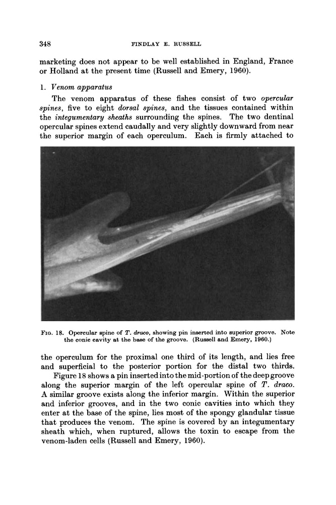
FIQ. 18. Opercular spine of T. draco, showing pin inserted into superior groove. Note
the conic cavity at the base of the groove. (Russell and Emery, 1960.)
the operculum for the proximal one third of its length, and lies free
and superficial to the posterior portion for the distal two thirds.
Figure 18 shows a pin inserted into the mid-portion of the deep groove
along the superior margin of the left opercular spine of T . draw.
A similar groove exists along the inferior margin. Within the superior
and inferior grooves, and in the two conic cavities into which they
enter a t the base of the spine, lies most of the spongy glandular tissue
that produces the venom. The spine is covered by an integumentary
sheath which, when ruptured, allows the toxin to escape from the
venom-laden cells (Russell and Emery, 1960).
FINDLAY E. RUSSELL
marketing does not appear to be well established in England, France
or Holland at the present time (Russell and Emery, 1960).
1. Venom apparatus
The venom apparatus of these fishes consist of two opercular
spines, five to eight dorsal spines, and the tissues contained within
the integumentary sheaths surrounding the spines. The two dentinal
opercular spines extend caudally and very slightly downward from near
the superior margin of each operculum. Each is firmly attached to
FIQ. 18. Opercular spine of T. draco, showing pin inserted into superior groove. Note
the conic cavity at the base of the groove. (Russell and Emery, 1960.)
the operculum for the proximal one third of its length, and lies free
and superficial to the posterior portion for the distal two thirds.
Figure 18 shows a pin inserted into the mid-portion of the deep groove
along the superior margin of the left opercular spine of T . draw.
A similar groove exists along the inferior margin. Within the superior
and inferior grooves, and in the two conic cavities into which they
enter a t the base of the spine, lies most of the spongy glandular tissue
that produces the venom. The spine is covered by an integumentary
sheath which, when ruptured, allows the toxin to escape from the
venom-laden cells (Russell and Emery, 1960).