

MARINE TOXINS AND VENOMOUS AND POISONOUS MARINE ANIMALS
341
Frequent, flattened, heavily pigmented cells are seen immediately
adjacent to the basal layer of the epithelium. In places these cells
form a distinct pigmented layer. The inner layer of basal cells is
perpendicularly oriented (stratified columnar of Ocampo et al., 1953).
As the basal cells mature their nuclei become smaller and stain more
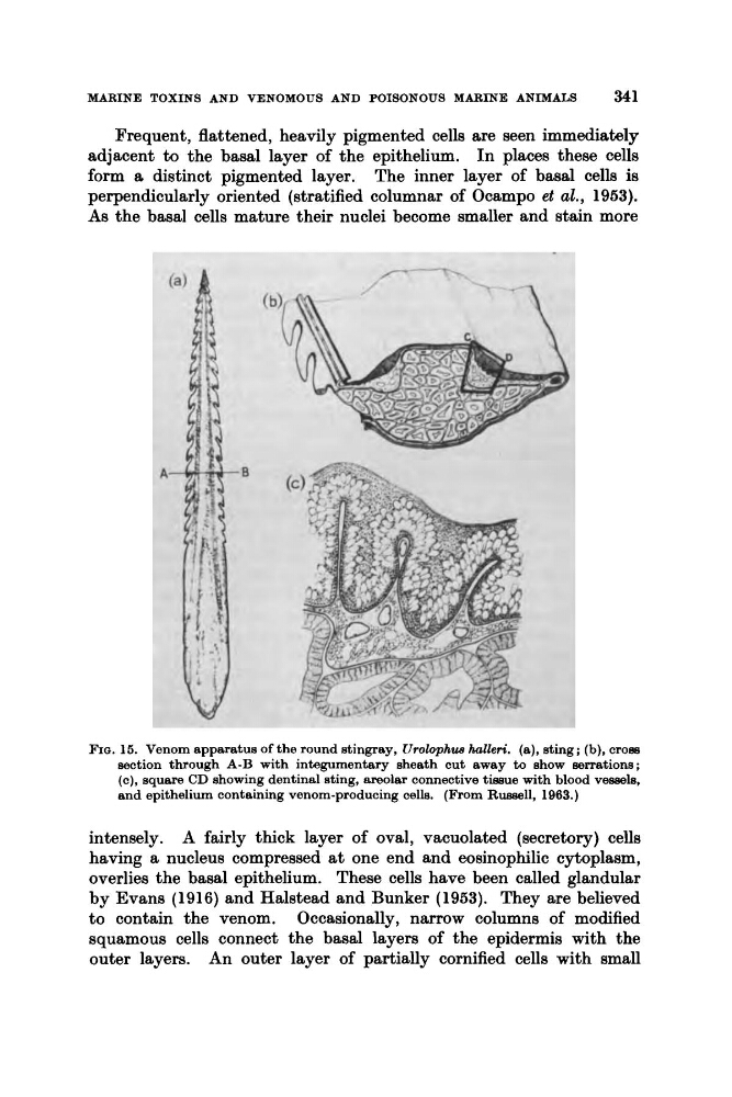
FIQ. 15. Venom apparatus of the round stingray, Urolophw halleri. (a), sting; (b), cross
section through A-B with integumentary sheath cut away to show serrations;
(c), square CD showing dentinal sting, areolar connective tissue with blood vessels,
and epithelium containing venom-producing cells. (From Russell, 1983.)
intensely. A fairly thick layer of oval, vacuolated (secretory) cells
having a nucleus compressed at one end and eosinophilic cytoplasm,
overlies the basal epithelium. These cells have been called glandular
by Evans (1916) and Halstead and Bunker (1953). They are believed
to contain the venom. Occasionally, narrow columns of modified
squamous cells connect the basal layers of the epidermis with the
outer layers. An outer layer of partially cornified cells with small
341
Frequent, flattened, heavily pigmented cells are seen immediately
adjacent to the basal layer of the epithelium. In places these cells
form a distinct pigmented layer. The inner layer of basal cells is
perpendicularly oriented (stratified columnar of Ocampo et al., 1953).
As the basal cells mature their nuclei become smaller and stain more
FIQ. 15. Venom apparatus of the round stingray, Urolophw halleri. (a), sting; (b), cross
section through A-B with integumentary sheath cut away to show serrations;
(c), square CD showing dentinal sting, areolar connective tissue with blood vessels,
and epithelium containing venom-producing cells. (From Russell, 1983.)
intensely. A fairly thick layer of oval, vacuolated (secretory) cells
having a nucleus compressed at one end and eosinophilic cytoplasm,
overlies the basal epithelium. These cells have been called glandular
by Evans (1916) and Halstead and Bunker (1953). They are believed
to contain the venom. Occasionally, narrow columns of modified
squamous cells connect the basal layers of the epidermis with the
outer layers. An outer layer of partially cornified cells with small