

LOCALIZATION IN THE DEVELOPING FUCUS EGG
315
•
• • •
m.
» * »
»»
Distance
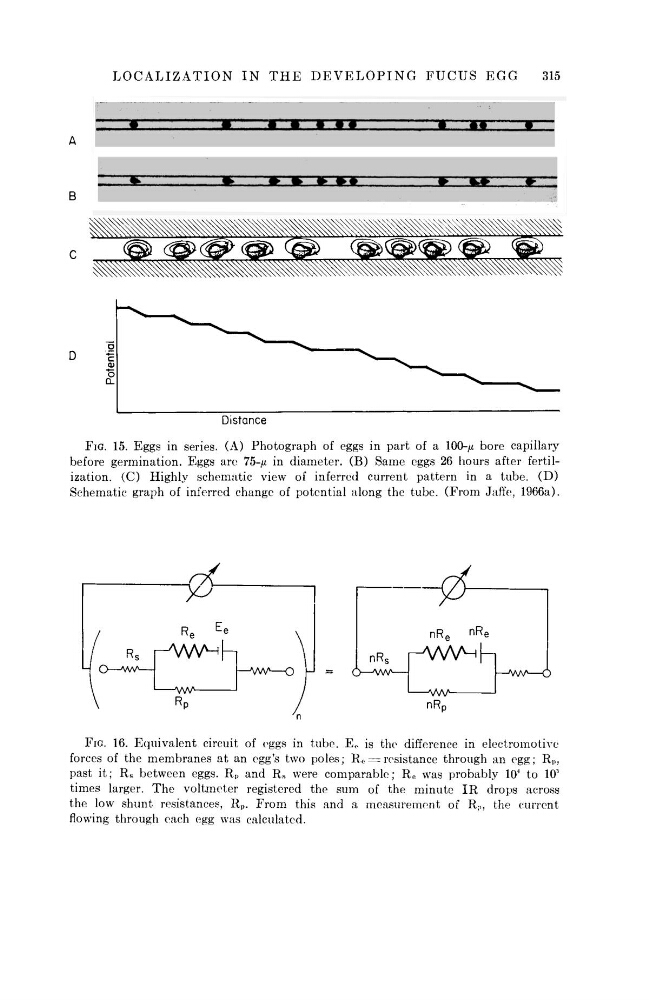
FIG. 15. Eggs in series. (A) Photograph of eggs in part of a 100-^ bore capillary
before germination. Eggs are 75-fj. in diameter. (B) Same eggs 26 hours after fertilization. (C) Highly schematic view of inferred current pattern in a tube. (D)
Schematic graph of inferred change of potential along the tube. (From Jaffe, 1966a).
at
R e
E
e
r w v ^ h
i—vw^Rp
Hvw—o
nR e
n R
e
nR s
r^VW^h
i
vW—w—6
nR D
FIG. 16. Equivalent circuit of eggs in tube. E 0 is the difference in electromotive
forces of the membranes at an egg's two poles; R e = resistance through an egg; R„,
past it; R s between eggs. R p and R s were comparable; R e was probably 10
4 to 10
5
times larger. The voltmeter registered the sum of the minute IR drops across
the low shunt resistances, R p . From this and a measurement of R ; ) , the current
flowing through each egg was calculated.
A
B
C
D
Potential
R s
r
-*AAA
315
•
• • •
m.
» * »
»»
Distance
FIG. 15. Eggs in series. (A) Photograph of eggs in part of a 100-^ bore capillary
before germination. Eggs are 75-fj. in diameter. (B) Same eggs 26 hours after fertilization. (C) Highly schematic view of inferred current pattern in a tube. (D)
Schematic graph of inferred change of potential along the tube. (From Jaffe, 1966a).
at
R e
E
e
r w v ^ h
i—vw^Rp
Hvw—o
nR e
n R
e
nR s
r^VW^h
i
vW—w—6
nR D
FIG. 16. Equivalent circuit of eggs in tube. E 0 is the difference in electromotive
forces of the membranes at an egg's two poles; R e = resistance through an egg; R„,
past it; R s between eggs. R p and R s were comparable; R e was probably 10
4 to 10
5
times larger. The voltmeter registered the sum of the minute IR drops across
the low shunt resistances, R p . From this and a measurement of R ; ) , the current
flowing through each egg was calculated.
A
B
C
D
Potential
R s
r
-*AAA