

D I F F E R E N T I A T I O N O F K I D N E Y M E S E N C H Y M E
273
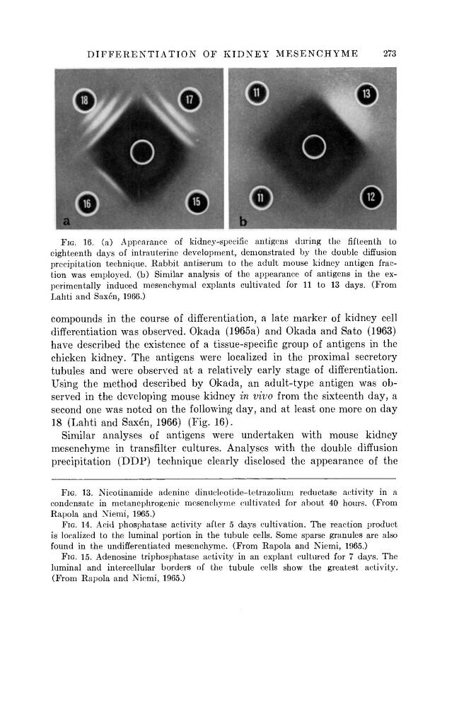
FIG. 16. (a) Appearance of kidney-specific antigens during the fifteenth to
eighteenth days of intrauterine development, demonstrated by the double diffusion
precipitation technique. Rabbit antiserum to the adult mouse kidney antigen fraction was employed, ( b ) Similar analysis of the appearance of antigens in the experimentally induced mesenchymal explants cultivated for 11 to 13 days. (From
Lahti and Saxen, 1966.)
compounds in the course of differentiation, a late marker of kidney cell
differentiation was observed. Okada (1965a) and Okada and Sato (1963)
have described the existence of a tissue-specific group of antigens in the
chicken kidney. T h e antigens were localized in the proximal secretory
tubules and were observed at a relatively early stage of differentiation.
Using the method described b y Okada, an adult-type antigen was o b -
served in the developing mouse kidney in vivo from the sixteenth day, a
second one was noted on the following d a y , and at least one more on d a y
18 (Lahti and Saxen, 1966) (Fig. 1 6 ) .
Similar analyses of antigens were undertaken with mouse kidney
mesenchyme in transfilter cultures. Analyses with the double diffusion
precipitation ( D D P ) technique clearly disclosed the appearance of the
FIG. 13. Nicotinamide adenine dinucleotide-tetrazolium reductase activity in a
condensate in metanephrogenic mesenchyme cultivated for about 40 hours. (From
Rapola and Niemi, 1965.)
FIG. 14. Acid phosphatase activity after 5 days cultivation. T h e reaction product
is localized to the luminal portion in the tubule cells. Some sparse granules are also
found in the undifferentiated mesenchyme. (From Rapola and Niemi, 1965.)
FIG. 15. Adenosine triphosphatase activity in an explant cultured for 7 days. The
luminal and intercellular borders of the tubule cells show the greatest activity.
(From Rapola and Niemi, 1965.)
273
FIG. 16. (a) Appearance of kidney-specific antigens during the fifteenth to
eighteenth days of intrauterine development, demonstrated by the double diffusion
precipitation technique. Rabbit antiserum to the adult mouse kidney antigen fraction was employed, ( b ) Similar analysis of the appearance of antigens in the experimentally induced mesenchymal explants cultivated for 11 to 13 days. (From
Lahti and Saxen, 1966.)
compounds in the course of differentiation, a late marker of kidney cell
differentiation was observed. Okada (1965a) and Okada and Sato (1963)
have described the existence of a tissue-specific group of antigens in the
chicken kidney. T h e antigens were localized in the proximal secretory
tubules and were observed at a relatively early stage of differentiation.
Using the method described b y Okada, an adult-type antigen was o b -
served in the developing mouse kidney in vivo from the sixteenth day, a
second one was noted on the following d a y , and at least one more on d a y
18 (Lahti and Saxen, 1966) (Fig. 1 6 ) .
Similar analyses of antigens were undertaken with mouse kidney
mesenchyme in transfilter cultures. Analyses with the double diffusion
precipitation ( D D P ) technique clearly disclosed the appearance of the
FIG. 13. Nicotinamide adenine dinucleotide-tetrazolium reductase activity in a
condensate in metanephrogenic mesenchyme cultivated for about 40 hours. (From
Rapola and Niemi, 1965.)
FIG. 14. Acid phosphatase activity after 5 days cultivation. T h e reaction product
is localized to the luminal portion in the tubule cells. Some sparse granules are also
found in the undifferentiated mesenchyme. (From Rapola and Niemi, 1965.)
FIG. 15. Adenosine triphosphatase activity in an explant cultured for 7 days. The
luminal and intercellular borders of the tubule cells show the greatest activity.
(From Rapola and Niemi, 1965.)