

GROWTH INDUCED BY DAMAGE
19
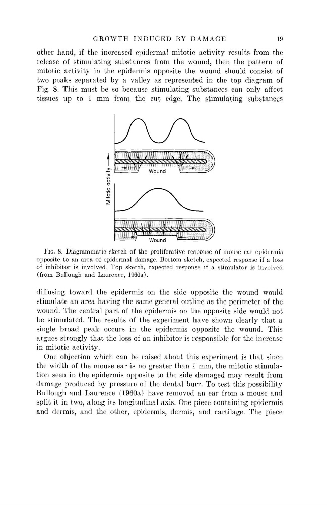
other hand, if the increased epidermal mitotic activity results from the
release of stimulating substances from the wound, then the pattern of
mitotic activity in the epidermis opposite the wound should consist of
two peaks separated by a valley as represented in the top diagram of
Fig. 8. This must be so because stimulating substances can only affect
tissues up to 1 mm from the cut edge. The stimulating substances
FIG. 8. Diagrammatic sketch of the proliferative response of mouse ear epidermis
opposite to an area of epidermal damage. Bottom sketch, expected response if a loss
of inhibitor is involved. Top sketch, expected response if a stimulator is involved
(from Bullough and Laurence, 1960a).
diffusing toward the epidermis on the side opposite the wound would
stimulate an area having the same general outline as the perimeter of the
wound. The central part of the epidermis on the opposite side would not
be stimulated. The results of the experiment have shown clearly that a
single broad peak occurs in the epidermis opposite the wound. This
argues strongly that the loss of an inhibitor is responsible for the increase
in mitotic activity.
One objection which can be raised about this experiment is that since
the width of the mouse ear is no greater than 1 mm, the mitotic stimulation seen in the epidermis opposite to the side damaged may result from
damage produced by pressure of the dental burr. To test this possibility
Bullough and Laurence (1960a) have removed an ear from a mouse and
split it in two, along its longitudinal axis. One piece containing epidermis
and dermis, and the other, epidermis, dermis, and cartilage. The piece
Mitotic activity —•
19
other hand, if the increased epidermal mitotic activity results from the
release of stimulating substances from the wound, then the pattern of
mitotic activity in the epidermis opposite the wound should consist of
two peaks separated by a valley as represented in the top diagram of
Fig. 8. This must be so because stimulating substances can only affect
tissues up to 1 mm from the cut edge. The stimulating substances
FIG. 8. Diagrammatic sketch of the proliferative response of mouse ear epidermis
opposite to an area of epidermal damage. Bottom sketch, expected response if a loss
of inhibitor is involved. Top sketch, expected response if a stimulator is involved
(from Bullough and Laurence, 1960a).
diffusing toward the epidermis on the side opposite the wound would
stimulate an area having the same general outline as the perimeter of the
wound. The central part of the epidermis on the opposite side would not
be stimulated. The results of the experiment have shown clearly that a
single broad peak occurs in the epidermis opposite the wound. This
argues strongly that the loss of an inhibitor is responsible for the increase
in mitotic activity.
One objection which can be raised about this experiment is that since
the width of the mouse ear is no greater than 1 mm, the mitotic stimulation seen in the epidermis opposite to the side damaged may result from
damage produced by pressure of the dental burr. To test this possibility
Bullough and Laurence (1960a) have removed an ear from a mouse and
split it in two, along its longitudinal axis. One piece containing epidermis
and dermis, and the other, epidermis, dermis, and cartilage. The piece
Mitotic activity —•